. 2019 Feb 14;15(2):e1007975.
doi: 10.1371/journal.pgen.1007975. eCollection 2019 Feb.

Assessing effects of germline exposure to environmental toxicants by high-throughput screening in C. elegans

Affiliations

Assessing effects of germline exposure to environmental toxicants by high-throughput screening in C. elegans

Nara Shin et al. PLoS Genet. .

Abstract

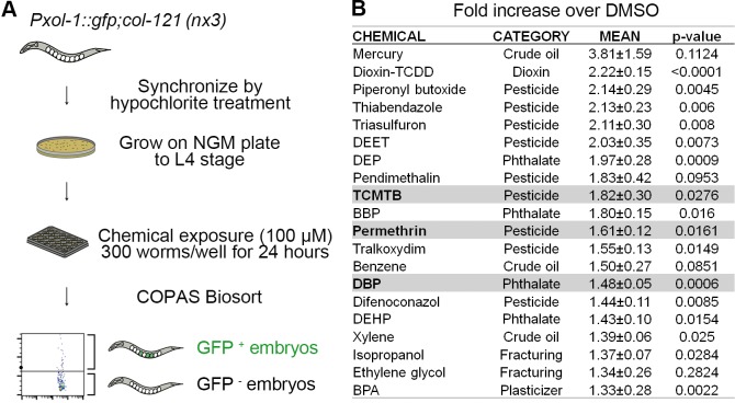
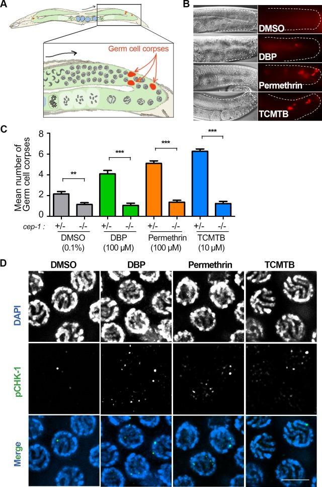
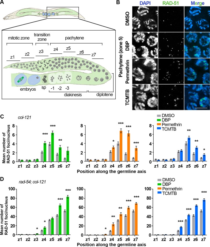
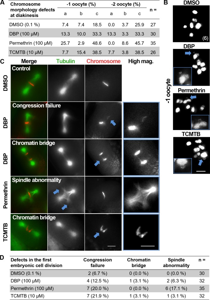
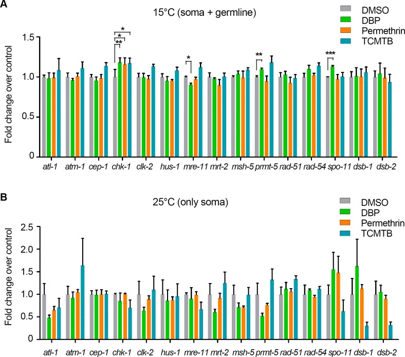
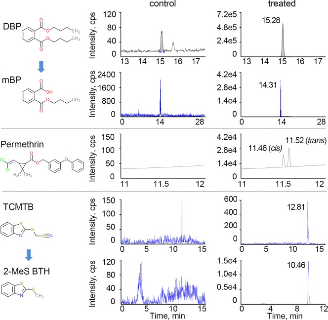
Chemicals that are highly prevalent in our environment, such as phthalates and pesticides, have been linked to problems associated with reproductive health. However, rapid assessment of their impact on reproductive health and understanding how they cause such deleterious effects, remain challenging due to their fast-growing numbers and the limitations of various current toxicity assessment model systems. Here, we performed a high-throughput screen in C. elegans to identify chemicals inducing aneuploidy as a result of impaired germline function. We screened 46 chemicals that are widely present in our environment, but for which effects in the germline remain poorly understood. These included pesticides, phthalates, and chemicals used in hydraulic fracturing and crude oil processing. Of the 46 chemicals tested, 41% exhibited levels of aneuploidy higher than those detected for bisphenol A (BPA), an endocrine disruptor shown to affect meiosis, at concentrations correlating well with mammalian reproductive endpoints. We further examined three candidates eliciting aneuploidy: dibutyl phthalate (DBP), a likely endocrine disruptor and frequently used plasticizer, and the pesticides 2-(thiocyanomethylthio) benzothiazole (TCMTB) and permethrin. Exposure to these chemicals resulted in increased embryonic lethality, elevated DNA double-strand break (DSB) formation, activation of p53/CEP-1-dependent germ cell apoptosis, chromosomal abnormalities in oocytes at diakinesis, impaired chromosome segregation during early embryogenesis, and germline-specific alterations in gene expression. This study indicates that this high-throughput screening system is highly reliable for the identification of environmental chemicals inducing aneuploidy, and provides new insights into the impact of exposure to three widely used chemicals on meiosis and germline function.


Conflict of interest statement

The authors have declared that no competing interests exist.

Figures

Fig 1
Fig 1. Flowchart for high-throughput screening strategy and readout.
(A) Worms carrying a collagen gene mutation (col-121(nx3)) for increased cuticle permeability and a male-specific promoter driving GFP expression in embryos (Pxol-1::gfp), were synchronized by hypochlorite treatment. Age-matched L1 stage animals were grown on plates (6,000 per 100 mm plate) until reaching the L4 stage. L4 stage animals were dispensed into 24-well plates (300 worms/well) with each well containing OP50 E. coli (OD600 = 24) in M9 buffer and a single chemical from our library (≤100 μM each) followed by a 24-hour incubation at 20°C. After thoroughly washed in M9, young adult animals were sorted based on fluorescence intensity with the COPAS Biosort. Adult worms with GFP+ embryos are detected as distinct from worms carrying GFP- embryos and debris, which are below the threshold (black horizontal line). Threshold was determined by comparing levels of GFP+ embryos detected in two genetic mutants, Pxol-1::gfp and Pxol-1::gfp;him-8. More than 5,000 animals were assessed in biological triplicates for each chemical exposure. (B) Readouts obtained with the COPAS Biosort for the chemicals that showed higher fold increase in GFP+ embryos compared to DMSO than BPA, a known endocrine disruptor (mean ± SEM; p-values calculated by the paired two-tailed t-test, 95% C.I.). The three chemicals highlighted in gray, TCTMB, permethrin, and DBP, were assessed further for their effects on germline functions.
Fig 2
Fig 2. DBP, Permethrin, and TCMTB exposures result in increased embryonic lethality, sterility and defects in chromosomal organization in the germline.
(A) Plate phenotypes for indicated chemical exposures. Embryonic lethality, larval lethality and the number of eggs laid (brood size) are shown for the indicated doses of exposures and compared to vehicle alone (0.1% DMSO). Error bars represent SEM. *P<0.05, **P<0.01, and ***P<0.001 by the two-tailed Mann-Whitney test, 95% C.I. (B) Images show pachytene nuclei in the germlines of whole, undissected worms fixed with Carnoy’s fixative and stained with DAPI that were exposed to vehicle control, 100 μM DBP, 100 μM permethrin, and 10 μM TCMTB. Chemical exposures lead to increased numbers of gonads with gaps (discontinuities or spaces lacking germ cell nuclei; arrow in second panel), aggregated nuclei (arrow, third panel) and DAPI-bright nuclei with chromosomes collapsed to one side (area indicated with a bracket, fourth panel) instead of dispersed throughout the nuclear periphery and organized in clear parallel tracks as seen in control at that stage. Scale bar, 5 μm. (C) Percentage of gonads exhibiting gaps, aggregates, or nuclei with chromatin in a leptotene/zygotene-like organization, in pachytene (n = number of gonads examined). * P<0.05, **P<0.01, and **P<0.001 by the two-sided Fisher’s exact test.
Fig 3
Fig 3. DBP, Permethrin, and TCMTB exposures lead to p53/CEP-1-dependent increased germ cell apoptosis and CHK-1 activation.
(A) Schematic representation of C. elegans and the area where DNA damage checkpoint activation of germ cell apoptosis is detected in the germline. Inset represents zoom-in of one gonad arm. Black arrow indicates orientation of progression through meiosis while red arrows indicate germ cell corpses (red) observed at late pachytene near the gonad bend. (B) Chemical exposures caused a significant increase in the number of germ cell corpses observed at late pachytene compared to vehicle alone. Gonads are traced to facilitate visualization. On the left is the Nomarski optics view and to the right are the acridine orange stained germ cell corpses (red). (C) Graphical representation showing mean number of germ cell corpses detected for each indicated chemical. Levels of germ cell corpses were significantly reduced in a p53/cep-1-dependent manner. Note that basal level of toxicity for DMSO, as previously described [42,103], is also reduced in a cep-1 mutant background. Analysis was done for three independent biological repeats. More than 30 gonads were scored for each chemical. Error bars represent SEM. **P<0.01, ***P<0.0001 by the two-tailed Mann-Whitney test, 95% C.I. (D) High-resolution images of mid to late pachytene nuclei from whole-mounted gonads immunostained for phospho CHK-1 (pCHK-1; green) and co-stained with DAPI (blue). Elevated levels of pCHK-1 were observed in chemical-treated worms compared to control. Scale bar, 5 μm.
Fig 4
Fig 4. Chemical exposures result in increased DSB formation and impaired DSB repair.
(A) Schematic representation of a C. elegans germline indicating the position of the equally sized zones (z1-z7) scored for RAD-51 foci. Nuclei in z1 and z2 are undergoing mitosis. They enter meiosis at z3 when they enter the transition zone, which corresponds to the leptotene/zygotene stages. Nuclei then proceed through pachytene (z4-z7), diplotene and diakinesis on their way into the uterus. sp: spermatheca. -1 indicates the oocyte closest to the spermatheca. (B) Representative images of pachytene nuclei (z5) immunostained for RAD-51 (green) and co-stained with DAPI (blue). Levels of RAD-51 foci in pachytene nuclei are elevated for each chemical exposure compared with control. Note that chromosomes in pachytene nuclei still exhibit a leptotene/zygotene-like organization in the cases of DBP and permethrin exposures. Scale bar, 5 μm. (C) Histograms show the mean number of RAD-51 foci scored per nucleus for each zone from col-121 worms. Elevated levels of foci are observed persisting until late pachytene indicating a defect in DSB repair. >5 gonads from three independent biological repeats were scored for each indicated exposure. Error bars represent SEM. (D) Quantification of the mean number of RAD-51 foci scored per nucleus in rad-54;col-121 worms. DSB levels were significantly higher upon the chemical exposures compared to vehicle alone. 3 gonads were scored for each chemical from two independent biological repeats. Error bars represent SEM. *P<0.05, **P<0.01, ***P<0.001 by the two-tailed Mann-Whitney test, 95% C.I.
Fig 5
Fig 5. Chemical exposures lead to defects at diakinesis and the first embryonic cell division.
(A) Quantification of the chromosome morphology defects observed in diakinesis (-1 and -2 oocytes). a: chromosome fragments, b: chromatin bridges, c: frayed chromosomes, n = total number of oocytes scored. (B) High resolution images of oocytes at diakinesis positioned right before the spermatheca (-1 oocyte). Six intact bivalents are observed in control. In contrast, frayed chromosomes, chromosome fragments and chromosome bridges (arrows and magnified in insets) are observed at higher levels in the germlines of chemical-exposed worms. Scale bar, 5 μm. (C) Representative images from time-lapse analysis of the first embryonic division in vehicle alone and DBP-, permethrin- and TCMTB-exposed H2B::mCherry; γ-tubulin::GFP; col-121(nx3) worms. A normal metaphase I configuration is shown for vehicle alone. Arrows and insets show examples of chromosomes that fail to align at the metaphase plate, chromatin bridges at the metaphase to anaphase transition and spindle abnormalities observed following exposures to all three chemicals. Scale bars, 5 μm. (D) Quantification of the time-lapse analysis of the first embryonic division. n = total number of embryos scored.
Fig 6
Fig 6. Germline-specific gene expression of DSB formation, repair and response genes is altered by chemical exposures.
Expression levels of a panel of genes responsible for DSB formation, repair and DNA damage response were examined by quantitative RT-PCR in glp-1(bn18);col-121(nx3) worms that grow without a germline when shifted from 15°C (A) to 25°C (B). Thus, 15°C represents gene expression in both soma and germline, while 25°C represents expression only in the soma. The expression levels of 15 critical and conserved genes were measured from either three (15°C) or four (25°C) independent biological replicates (each performed with technical triplicates) and normalized to gpd-1 (GAPDH). Y-axis indicates gene expression level change relative to vehicle alone. Error bars represent SEM. *P<0.05, **P<0.01, ***P<0.001 by the unpaired two-tailed t-test.
Fig 7
Fig 7. Structures and representative LC-MS/MS and GC-MS chromatograms for DBP, permethrin, TCMTB and their metabolites.
Chromatograms on the left show results from the analysis of the indicated chemicals and metabolites in worms exposed to vehicle alone (DMSO; control) and chromatograms on the right show results from worms exposed to DBP, permethrin, and TCMTB (treated). These chemicals were unambiguously identified and quantified using their respective internal standards. Y-axis represents relative abundance of signal intensity and X-axis represents retention time in minutes. The detected concentrations following chemical treatments (right) were consistently greater than in control (note differences in Y-axis). Note that the peak at 12.81 minutes does not correspond to TCMTB (expected peak for TCMTB is at 8.76), suggesting that TCMTB is very rapidly metabolized in the worm.

Similar articles

Cited by

References

    1. Siracusa JS, Yin L, Measel E, Liang S, Yu X. Effects of bisphenol A and its analogs on reproductive health: A mini review. Reprod Toxicol. 2018;79: 96–123. 10.1016/j.reprotox.2018.06.005 - DOI - PMC - PubMed
    1. Noorimotlagh Z, Haghighi NJ, Ahmadimoghadam M, Rahim F. An updated systematic review on the possible effect of nonylphenol on male fertility. EnvironSci -Pollut Res. 2017;24: 3298–3314. 10.1007/s11356-016-7960-y - DOI - PubMed
    1. Tomza-Marciniak A, Stępkowska P, Kuba J, Pilarczyk B. Effect of bisphenol A on reproductive processes: A review of in vitro, in vivo and epidemiological studies: Effect of BPA on reproductive processes. J Appl Toxicol. 2018;38: 51–80. 10.1002/jat.3480 - DOI - PubMed
    1. Del-Mazo J, Brieño-Enríquez MA, García-López J, López-Fernández LA, De-Felici M. Endocrine disruptors, gene deregulation and male germ cell tumors. IntJ Dev Biol. 2013;57: 225–239. 10.1387/ijdb.130042jd - DOI - PubMed
    1. Brieño-Enríquez MA, Larriba E, del Mazo J. Endocrine disrupters, microRNAs, and primordial germ cells: a dangerous cocktail. Fertil Steril. 2016;106: 871–879. 10.1016/j.fertnstert.2016.07.1100 - DOI - PubMed

Publication types

MeSH terms