

A chronic bioluminescent model of experimental visceral leishmaniasis for accelerating drug discovery
- PMID: 30763330
- PMCID: PMC6392311
- DOI: 10.1371/journal.pntd.0007133
A chronic bioluminescent model of experimental visceral leishmaniasis for accelerating drug discovery
Abstract
Background: Visceral leishmaniasis is a neglected parasitic disease with no vaccine available and its pharmacological treatment is reduced to a limited number of unsafe drugs. The scarce readiness of new antileishmanial drugs is even more alarming when relapses appear or the occurrence of hard-to-treat resistant strains is detected. In addition, there is a gap between the initial and late stages of drug development, which greatly delays the selection of leads for subsequent studies.
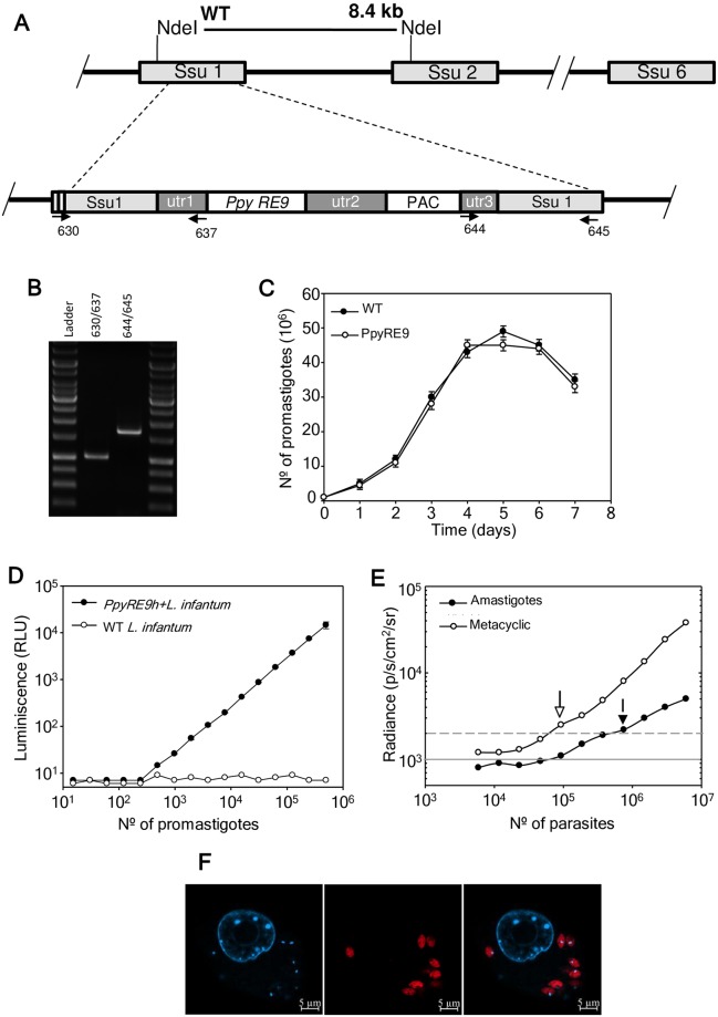
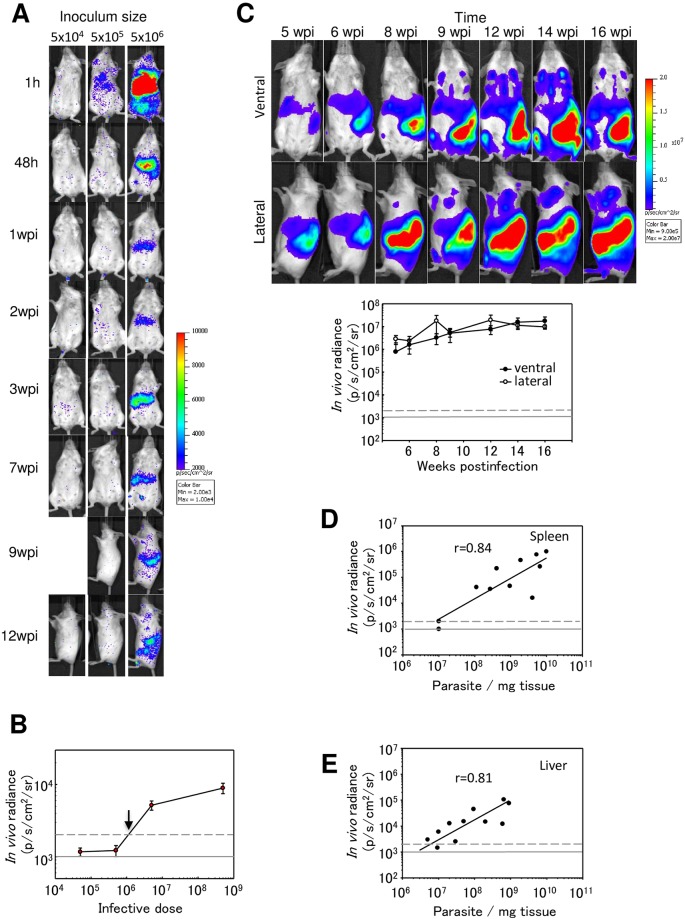
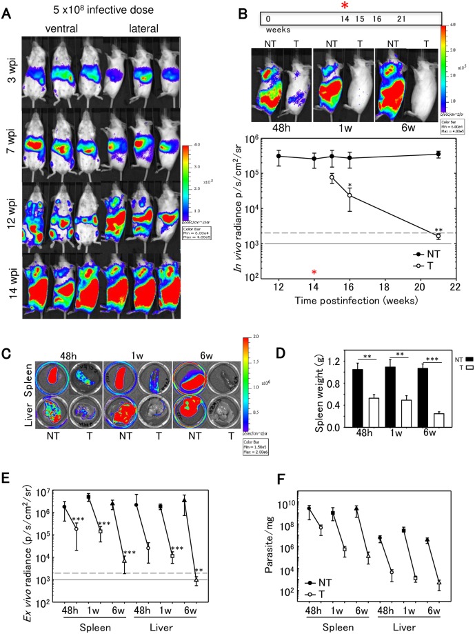
Methodology/principal findings: In order to address these issues, we have generated a red-shifted luminescent Leishmania infantum strain that enables long-term monitoring of parasite burden in individual animals with an in vivo limit of detection of 106 intracellular amastigotes 48 h postinfection. For this purpose, we have injected intravenously different infective doses (104-5x10⁸) of metacyclic parasites in susceptible mouse models and the disease was monitored from initial times to 21 weeks postinfection. The emission of light from the target organs demonstrated the sequential parasite colonization of liver, spleen and bone marrow. When miltefosine was used as proof-of-concept, spleen weight parasite burden and bioluminescence values decreased significantly.
Conclusions: In vivo bioimaging using a red-shifted modified Leishmania infantum strain allows the appraisal of acute and chronic stage of infection, being a powerful tool for accelerating drug development against visceral leishmaniasis during both stages and helping to bridge the gap between early discovery process and subsequent drug development.
Conflict of interest statement
The authors have declared that no competing interests exist.
Figures
Similar articles
-
Generation of luciferase-expressing Leishmania infantum chagasi and assessment of miltefosine efficacy in infected hamsters through bioimaging.PLoS Negl Trop Dis. 2015 Feb 13;9(2):e0003556. doi: 10.1371/journal.pntd.0003556. eCollection 2015 Feb. PLoS Negl Trop Dis. 2015. PMID: 25679212 Free PMC article.
-
Evidence of a drug-specific impact of experimentally selected paromomycin and miltefosine resistance on parasite fitness in Leishmania infantum.J Antimicrob Chemother. 2016 Jul;71(7):1914-21. doi: 10.1093/jac/dkw096. Epub 2016 Apr 15. J Antimicrob Chemother. 2016. PMID: 27084919
-
Potentiation of the leishmanicidal activity of nelfinavir in combination with miltefosine or amphotericin B.Int J Antimicrob Agents. 2018 Nov;52(5):682-687. doi: 10.1016/j.ijantimicag.2018.06.016. Epub 2018 Jun 30. Int J Antimicrob Agents. 2018. PMID: 29969693
-
The preclinical discovery and development of oral miltefosine for the treatment of visceral leishmaniasis: a case history.Expert Opin Drug Discov. 2020 Jun;15(6):647-658. doi: 10.1080/17460441.2020.1743674. Epub 2020 Mar 23. Expert Opin Drug Discov. 2020. PMID: 32202449 Review.
-
Miltefosine--discovery of the antileishmanial activity of phospholipid derivatives.Trans R Soc Trop Med Hyg. 2006 Dec;100 Suppl 1:S4-8. doi: 10.1016/j.trstmh.2006.03.009. Epub 2006 Aug 14. Trans R Soc Trop Med Hyg. 2006. PMID: 16904717 Review.
Cited by
-
Resistance to Experimental Visceral Leishmaniasis in Mice Infected With Leishmania infantum Requires Batf3.Front Immunol. 2020 Dec 10;11:590934. doi: 10.3389/fimmu.2020.590934. eCollection 2020. Front Immunol. 2020. PMID: 33362772 Free PMC article.
-
Screening Marine Natural Products for New Drug Leads against Trypanosomatids and Malaria.Mar Drugs. 2020 Mar 31;18(4):187. doi: 10.3390/md18040187. Mar Drugs. 2020. PMID: 32244488 Free PMC article. Review.
-
Generation and Characterization of a Dual-Reporter Transgenic Leishmania braziliensis Line Expressing eGFP and Luciferase.Front Cell Infect Microbiol. 2020 Jan 22;9:468. doi: 10.3389/fcimb.2019.00468. eCollection 2019. Front Cell Infect Microbiol. 2020. PMID: 32039047 Free PMC article.
-
Systematic identification of genes encoding cell surface and secreted proteins that are essential for in vitro growth and infection in Leishmania donovani.PLoS Pathog. 2022 Feb 24;18(2):e1010364. doi: 10.1371/journal.ppat.1010364. eCollection 2022 Feb. PLoS Pathog. 2022. PMID: 35202447 Free PMC article.
-
Establishment, optimisation and quantitation of a bioluminescent murine infection model of visceral leishmaniasis for systematic vaccine screening.Sci Rep. 2020 Mar 13;10(1):4689. doi: 10.1038/s41598-020-61662-3. Sci Rep. 2020. PMID: 32170135 Free PMC article.
References
-
- Pace D. Leishmaniasis. J Infect. 2014;69 Suppl 1:S10–18. - PubMed
