. 2019 Feb 14;14(2):e0211904.
doi: 10.1371/journal.pone.0211904. eCollection 2019.

TyrR is involved in the transcriptional regulation of biofilm formation and D-alanine catabolism in Azospirillum brasilense Sp7

Affiliations

TyrR is involved in the transcriptional regulation of biofilm formation and D-alanine catabolism in Azospirillum brasilense Sp7

Saúl Jijón-Moreno et al. PLoS One. .

Abstract

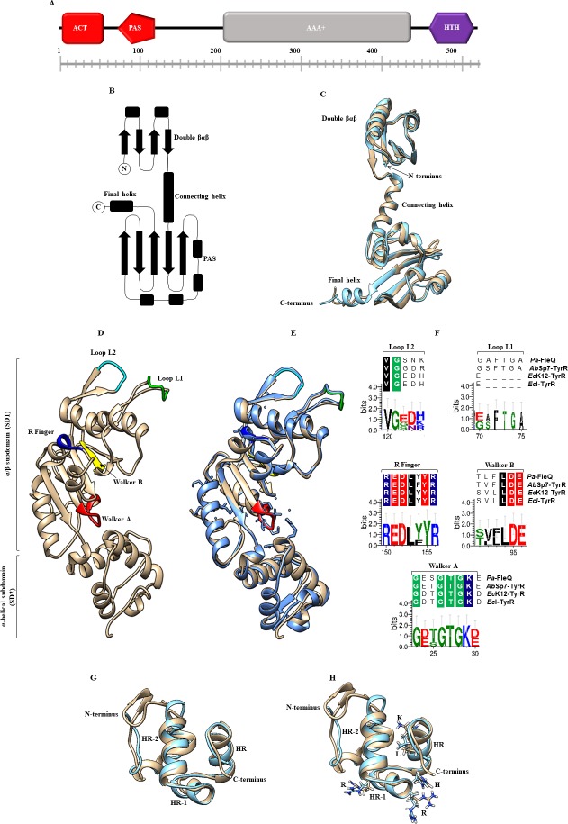
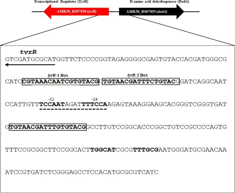
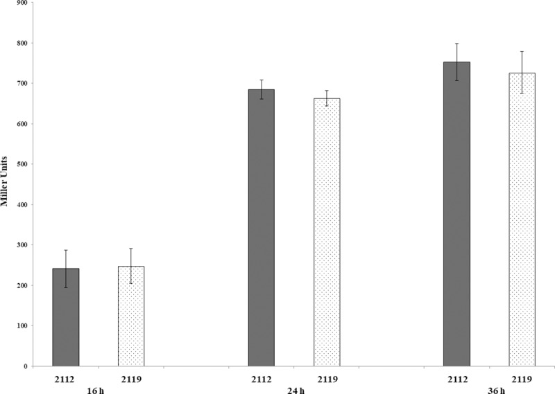
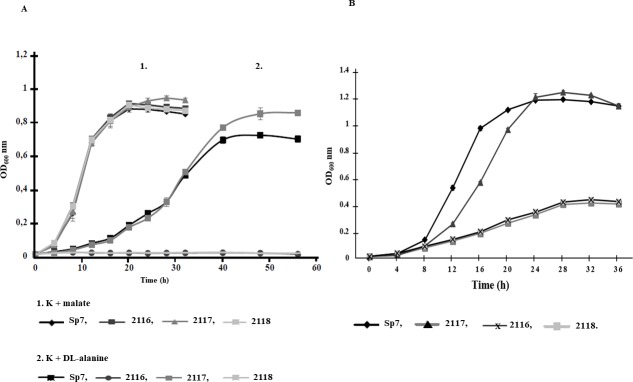
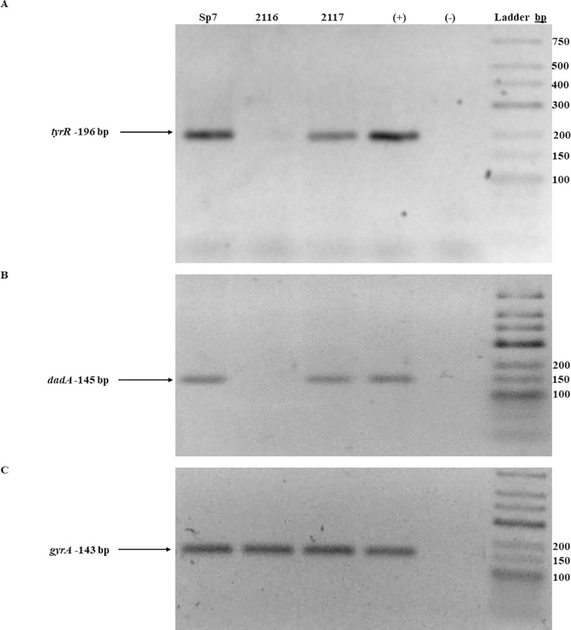
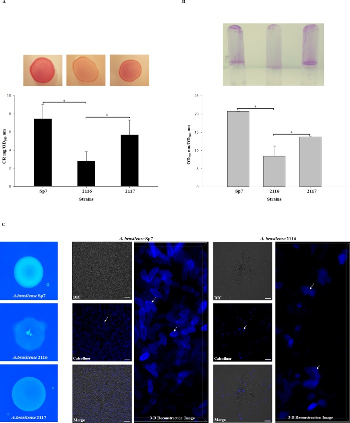
Azospirillum brasilense is one of the most studied species of diverse agronomic plants worldwide. The benefits conferred to plants inoculated with Azospirillum have been primarily attributed to its capacity to fix atmospheric nitrogen and synthesize phytohormones, especially indole-3-acetic acid (IAA). The principal pathway for IAA synthesis involves the intermediate metabolite indole pyruvic acid. Successful colonization of plants by Azospirillum species is fundamental to the ability of these bacteria to promote the beneficial effects observed in plants. Biofilm formation is an essential step in this process and involves interactions with the host plant. In this study, the tyrR gene was cloned, and the translated product was observed to exhibit homology to TyrR protein, a NtrC/NifA-type activator. Structural studies of TyrR identified three putative domains, including a domain containing binding sites for aromatic amino acids in the N-terminus, a central AAA+ ATPase domain, and a helix-turn-helix DNA binding motif domain in the C-terminus, which binds DNA sequences in promoter-operator regions. In addition, a bioinformatic analysis of promoter sequences in A. brasilense Sp7 genome revealed that putative promoters encompass one to three TyrR boxes in genes predicted to be regulated by TyrR. To gain insight into the phenotypes regulated by TyrR, a tyrR-deficient strain derived from A. brasilense Sp7, named A. brasilense 2116 and a complemented 2116 strain harboring a plasmid carrying the tyrR gene were constructed. The observed phenotypes indicated that the putative transcriptional regulator TyrR is involved in biofilm production and is responsible for regulating the utilization of D-alanine as carbon source. In addition, TyrR was observed to be absolutely required for transcriptional regulation of the gene dadA encoding a D-amino acid dehydrogenase. The data suggested that TyrR may play a major role in the regulation of genes encoding a glucosyl transferase, essential signaling proteins, and amino acids transporters.


Conflict of interest statement

The authors have declared that no competing interests exist.

Figures

Fig 1
Fig 1. The overall predicted and domains displayed by the TyrR protein from A. brasilense Sp7 (Ab-Sp7).
(A) The TyrR protein consists of an ACT domain (red square), a PAS domain (red pentagon), an AAA+ domain (gray rectangle) and an HTH motif (purple hexagon). (B) The topology of the N-terminal region of a TyrR monomer from E. coli K12 with its separate domains. (C) Comparison by superposition of monomers of TyrR from the E. coli K12 (blue) crystal structure with a resolution of 2.3 Å and Ab-Sp7 (beige) with a root mean square deviation (RMSD) of 1.59 Å. (D) The prediction of the AAA+ monomer domain of TyrR from Ab-Sp7. The ribbon representation of Ab_TyrR-AAA+ consists of an α/β sub domain followed by a smaller α-helical subdomain. The regions and the PAS domain are highlighted. (E) Superimposition of the Pa_FleQ-AAA+ crystal structure with a resolution of 1.8 Å (blue) and Ab_TyrR-AAA+ (beige) with a RSMD of 0.89 Å. The significant motifs are highlighted: Walker A (red), Walker B (yellow), R finger (dark blue), Loop L1, (green), Loop L2 (cyan). (F) Sequence logo representations of sequence alignments of Walker A (GXXXXGK), Walker B (hhhhDE), R finger (RXXXXXR), Loop L2 (VG), Loop L1 (GAFTGA), and multiple sequence alignments of FleQ (P. aeruginosa), TyrR (A. brasilense Sp7), TyrR (E. coli K12), TyrR (E. cloacae) were used. (G) Comparison by structure superimposition of the Ab-TyrR-HTH model (beige) and template 1G2H of H. influenzae (blue color). Three well defined α-helices (HR-2, HR-1, and HR) are shown. (H) Superimposed structures show the crucial amino acids involved in forming hydrogen bonds directly with DNA are highlighted: arginine 291 (R), arginine 296 (R), histidine 301 (H), lysine 307 (K), and leucine 308 (L). The homology models constructed by I-Tasser with the best scores, C-score -1.21, RMSD 1.25 Å, visualized with UCSF Chimera software. Logos were generated with the Weblogo 3 Create software.
Fig 2
Fig 2. Sequences of the cis elements contained in the intergenic promoter region of the tyrR and dadA genes from A. brasilense Sp7.
The TyrR boxes are shown in blod and are included in a rectangle; the -12 and -24 promoter regions are indicated in bold and with lines. The arrows indicate the ORFs of the tyrR and dadA genes. Identification of tyrR boxes in the A. brasilense Sp7 genome using the Find Individual Motif Occurrences (FIMO) software tool, which is part of the MEME Suite software toolkit [42].
Fig 3
Fig 3. Expression analysis of ipdC-lacZ transcriptional fusion under the control of A. brasilense 2119 tyrR minus strain.
A. brasilense strains were grown in minimal media (MM) with malate, as described in the Material and Methods section, during 16, 24 and 36 h. Data are reported as Miller units by mg of protein from three independent experiments with two biological replicates.
Fig 4
Fig 4. Growth profiles of the A. brasilense Sp7, 2116, 2117, and 2118 strains.
(A) Growth kinetics of A. brasilense strains in minimal media (MM) with malate or D-alanine as the sole carbon sources. The strains were grown in MM supplemented with malate (20 mM) or DL-alanine (20 mM) as carbon sources. Growth was followed at OD600 nm every 4 h until 56 h. Sp7, 2116, 2117 and 2118 strains.The data points represent the average from three independent cultures of each strain. (B) Growth of A. brasilense strains in minimal medium with D-alanine as the sole nitrogen source. Bacterial strains were grown in DL-malate minimal medium supplemented with D-alanine (20 mM) as the sole nitrogen source. Growth was followed at OD600 nm every 4 h until 36 h. The data points represent the average from three independent cultures of each strain.
Fig 5
Fig 5. RT-PCR analysis of dadA, tyrR and gyrA expression from the A. brasilense Sp7, 2116, and 2117 strains.
RT-PCR analysis of tyrR, dadA and gyrA expression from the A. brasilense Sp7, 2116 and 2117 strains. Total RNA (1 μg) from each strain was subjected to reverse transcription, and the obtained cDNA was amplified using pairs of primers specific for tyrR. (A) dadA, (B), and gyrA (C) A. brasilense strains are indicated at the top of each lane by their conventional denomination. The expected PCR products of 196 bp (tyrR), 145 bp (dadA) and 143 bp (gyrA) were visualized by ethidium bromide staining on a 2.5% agarose gel. A positive control (+), represented by genomic DNA. A negative reaction control is represented by (-), without reverse transcriptase, both were included in each experiment. The image is representative of three independents experiments.
Fig 6
Fig 6. Biofilms and EPS production by the A. brasilense Sp7, 2116, and 2117 strains.
(A) CR staining assay. Cells were grown on agar-solidified CR medium inoculated with 10 μL of 1.0 OD600 nm of each culture strains for 72 h at 30 ºC. For EPS quantification, the strains were cultured as described in Material and Methods and incubated at 30°C for 5 days. CR binding was expressed as mg CR/OD600 nm. (B) For biofilm production cells were grown statically for 5 days at 30 ºC on NFb* with 27mM malate as a C source + 10mM D-alanine. Biofilm formation was visualized by Crystal violet staining as well quantified and normalized per OD600 nm of growth. All data are the average from three independent experiments performed in duplicate. Error bars indicate standard errors of the means as compared with values observed for WT. For all data, P was < 0.001 as assessed by Student´s t-test. (C) Calcofluor white colorant M2R (CWC) staining. Cells were grown on agar-solidified NFb* supplemented with CWC and inoculated with 10 μL of 1.0 OD600 nm of each culture strains for five days at 30 ºC. The cell fluorescence was observed under was observed with a Nikon Eclipse Ti-E C2+. Bars represent 10 μm. The white arrows indicate EPS.

Similar articles

Cited by

References

    1. Argaet VP, Wilson TJ, Davidson BE. Purification of the Escherichia coli regulatory protein TyrR and analysis of its interactions with ATP, tyrosine, phenylalanine, and tryptophan. The Journal of biological chemistry. 1994;269(7):5171–8. . - PubMed
    1. Pittard J, Camakaris H, Yang J. The TyrR regulon. Mol Microbiol. 2005;55(1):16–26. 10.1111/j.1365-2958.2004.04385.x WOS:000225949300003. - DOI - PubMed
    1. Yang J, Ogawa Y, Camakaris H, Shimada T, Ishihama A, Pittard AJ. folA, a new member of the TyrR regulon in Escherichia coli K-12. Journal of bacteriology. 2007;189(16):6080–4. 10.1128/JB.00482-07 - DOI - PMC - PubMed
    1. Yang J, Hwang JS, Camakaris H, Irawaty W, Ishihama A, Pittard J. Mode of action of the TyrR protein: repression and activation of the tyrP promoter of Escherichia coli. Mol Microbiol. 2004;52(1):243–56. 10.1111/j.1365-2958.2003.03965.x . - DOI - PubMed
    1. Smith HQ, Somerville RL. The tpl promoter of Citrobacter freundii is activated by the TyrR protein. Journal of bacteriology. 1997;179(18):5914–21. 10.1128/jb.179.18.5914-5921.1997 WOS:A1997XV69900035. - DOI - PMC - PubMed

Publication types

MeSH terms