. 2019 Feb 14;13(2):e0007197.
doi: 10.1371/journal.pntd.0007197. eCollection 2019 Feb.

Renal and vascular effects of kallikrein inhibition in a model of Lonomia obliqua venom-induced acute kidney injury

Affiliations

Renal and vascular effects of kallikrein inhibition in a model of Lonomia obliqua venom-induced acute kidney injury

Markus Berger et al. PLoS Negl Trop Dis. .

Abstract

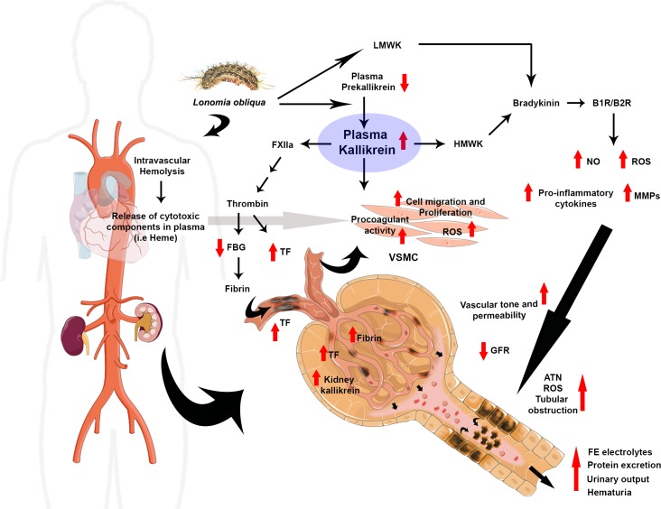
Background: Lonomia obliqua venom is nephrotoxic and acute kidney injury (AKI) is the main cause of death among envenomed victims. Mechanism underlying L. obliqua-induced AKI involves renal hypoperfusion, inflammation, tubular necrosis and loss of glomerular filtration and tubular reabsorption capacities. In the present study, we aimed to investigate the contribution of kallikrein to the hemodynamic instability, inflammation and consequent renal and vascular impairment.

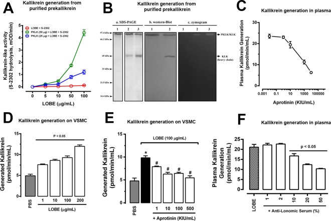
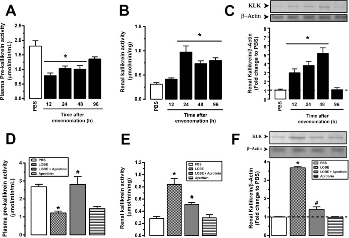
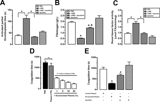
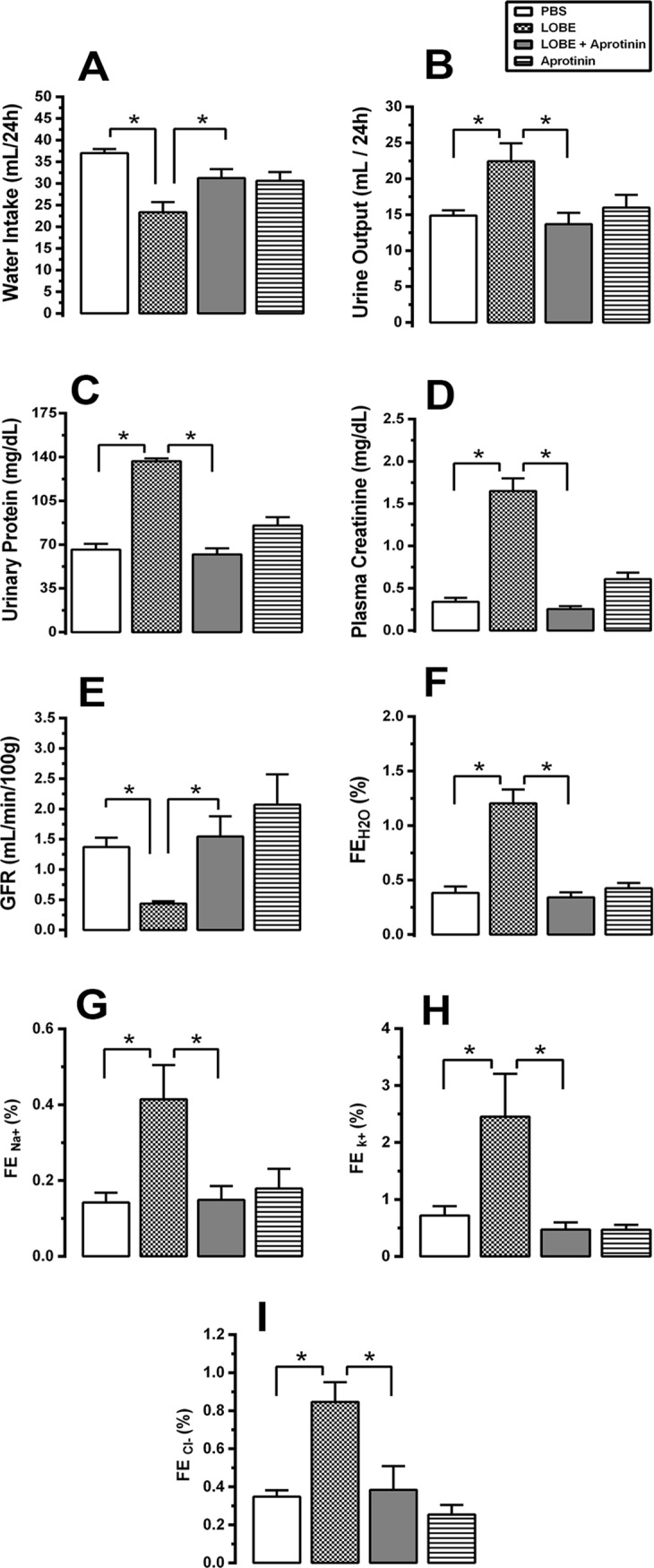
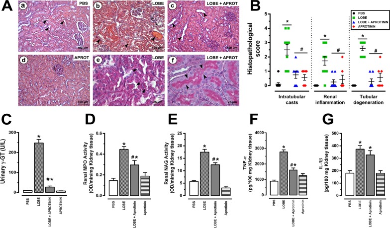
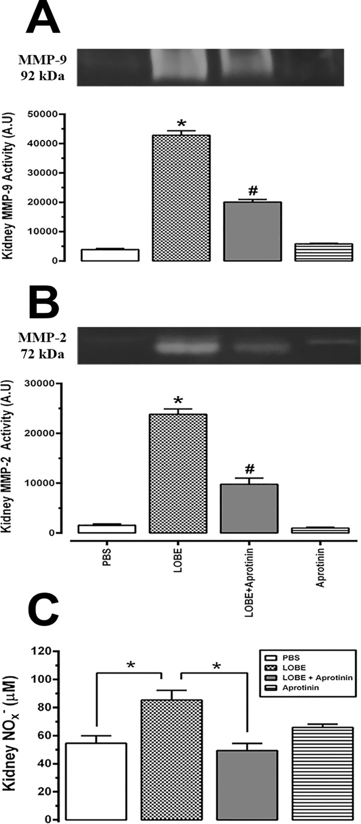
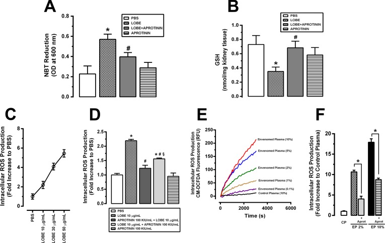
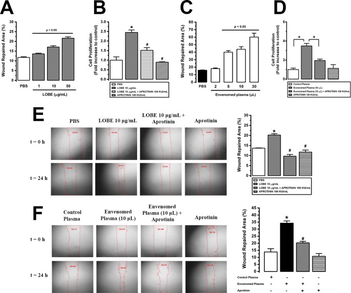
Methodology/principal findings: Addition of L. obliqua venom to purified prekallikrein and human plasma in vitro or to vascular smooth muscle cells (VSMC) in culture, was able to generate kallikrein in a dose-dependent manner. Injected in rats, the venom induced AKI and increased kallikrein levels in plasma and kidney. Kallikrein inhibition by aprotinin prevented glomerular injury and the decrease in glomerular filtration rate, restoring fluid and electrolyte homeostasis. The mechanism underlying these effects was associated to lowering renal inflammation, with decrease in pro-inflammatory cytokines and matrix metalloproteinase expression, reduced tubular degeneration, and protection against oxidative stress. Supporting the key role of kallikrein, we demonstrated that aprotinin inhibited effects directly associated with vascular injury, such as the generation of intracellular reactive oxygen species (ROS) and migration of VSMC induced by L. obliqua venom or by diluted plasma obtained from envenomed rats. In addition, kallikrein inhibition also ameliorated venom-induced blood incoagulability and decreased kidney tissue factor expression.

Conclusions/significance: These data indicated that kallikrein and consequently kinin release have a key role in kidney injury and vascular remodeling. Thus, blocking kallikrein may be a therapeutic alternative to control the progression of venom-induced AKI and vascular disturbances.


Conflict of interest statement

The authors have declared that no competing interests exist.

Figures

Fig 1
Fig 1. L. obliqua venom-induced prekallikrein activation.
A. Different concentrations of L. obliqua bristle extract (LOBE) was incubated in the presence or absence of purified plasma prekallikrein (PKLK) and generated kallikrein (KLK) was then measured by the addition of S-2302. B. LOBE-induced PKLK activation was confirmed by SDS-PAGE, western-blot and zymogram gels. a. SDS-PAGE: 1 –purified plasma PKLK (25 μg); 2 –Purified PKLK (25 μg) previously incubated with LOBE (5 μg) during 15 min; 3—Purified PKLK (25 μg) previously incubated with LOBE (5 μg) during 30 min. All samples were running in 12% gels under reducing conditions. b. western-blot: 1 –Purified PKLK (25 μg); 2—Purified PKLK (25 μg) previously incubated with LOBE (5 μg) during 30 min. In both cases, samples were running under reducing conditions and plasma PKLK or KLK heavy chain were identified using a specific anti-plasma PKLK antibody. c. zymogram. 1 –purified plasma PKLK (25 μg); 2 –Purified PKLK (25 μg) previously incubated with LOBE (5 μg) during 15 min; 3—Purified PKLK (25 μg) previously incubated with LOBE (5 μg) during 30 min. The enzymatic activity of generated KLK was detected by SDS-PAGE 12% containing a mixture of casein + plasminogen under non-reducing conditions. C. Human plasma was incubated with different concentrations of aprotinin and kallikrein generation was triggered by the addition of LOBE (100 μg/mL). D. VSMC culture were treated with different concentrations of LOBE for 24h and washed twice in PBS. Then the monolayers were incubated with human plasma and contact system activation was monitored through kallikrein formation. E. VSMC culture were treated (24h) with different concentrations of aprotinin, washed twice, and treated with LOBE (100 μg/mL) for 6h. After a second step wash, VSMC were incubated with human plasma and kallikrein formation was monitored. F. Human plasma was incubated with different concentrations of anti-lonomic serum (anti-venom produced by the Butantan Institute in Brazil) and kallikrein generation was triggered by the addition of LOBE (100 μg/mL). In all experiments the kinetics of kallikrein generation was monitored using the chromogenic substrate S-2302. Data represents mean of three independent experiments ± SE. * denotes p<0.05 vs PBS group and # denotes p<0.05 vs group treated with LOBE (100 μg/mL). KIU denotes kallikrein inhibitory units.
Fig 2
Fig 2. Kallikrein is generated during L. obliqua envenomation in vivo.
Wistar rats were injected with LOBE (1.5 mg/kg, s.c) and the following parameters were determined at different times post-envenomation: A. Plasma prekallikrein levels. B. Renal kallikrein activity. C. Protein levels of renal kallikrein determined by western-blot using specific antibody against tissue kallikrein. In another set of experiments, groups of animals were treated with PBS, LOBE (1.5 mg/kg, s.c), aprotinin (40,000 KIU/mg, i.v) or received aprotinin (40,000 KIU/mg, i.v) 30 min prior to the injection of the LOBE (1.5 mg/kg, sc). After 24h the same parameters were determined: D. Plasma prekallikrein levels. E. Renal kallikrein activity. F. Protein levels of renal tissue kallikrein. Data represents mean of three independent experiments ± SE. * denotes p<0.05 vs PBS group and # denotes p<0.05 vs group treated with LOBE.
Fig 3
Fig 3. Kallikrein generated during L. obliqua envenomation in vivo is directly involved in venom-induced coagulation disturbances.
Wistar rats were treated with PBS, LOBE (1.5 mg/kg, s.c), aprotinin (40,000 KIU/mg, i.v) or received aprotinin (40,000 KIU/mg, i.v) 30 min prior to the injection of LOBE (1.5 mg/kg, sc). After 24h the following renal parameters were determined: A. Activated partial thromboplastin time (APTT). B. Fibrinogen levels. C. Renal tissue factor levels. D. VSMC were cultured in the presence of plasma from control (animals treated with PBS) or envenomed rats. After 24h the monolayers were washed twice and coagulation time was determined by adding normal rat plasma in the presence of calcium ions. E. VSMC culture were treated (24h) with aprotinin (100 KIU/mL), washed twice and then treated with envenomed or control plasma (10%) for 6h. After a second step wash, VSMC were incubated with normal rat plasma and coagulation time was determined. Data represents mean ± SE. * denotes p<0.05 vs PBS group and # denotes p<0.05 vs group treated with LOBE or envenomed plasma; CTRL denotes control and ns denotes difference not significant.
Fig 4
Fig 4. Kallikrein activation during L. obliqua envenomation plays a central role in renal functional impairment.
Wistar rats were treated with PBS, LOBE (1.5 mg/kg, s.c), aprotinin (40,000 KIU/mg, i.v) or received aprotinin (40,000 KIU/mg, i.v) 30 min prior to the injection of LOBE (1.5 mg/kg, sc). After 24h the following renal parameters were determined: A. Water intake; B. Urine output; C. Urinary protein; D. Plasma creatinine; E. Glomerular filtration rate (GFR); F. Fractional excretion of water (FEH2O); G. Fractional excretion of sodium (FENa+); H. Fractional excretion of potassium (FEK+) and I. Fractional excretion of chloride (FECl-). Data represents mean ± SE. * denotes p<0.05.
Fig 5
Fig 5. Effects of aprotinin on renal inflammation, tubular degeneration and morphological alterations induced by L. obliqua venom.
A. Kidney morphological changes of Wistar rats pre-treated or not with aprotinin after 24h of envenomation. (a) cortical section stained with HE of an animal injected with PBS, showing normal glomerular and tubular (arrowheads) structures; (b) cortical section stained with HE of an animal injected with LOBE (1.5 mg/kg, s.c), showing acute tubular necrosis, intratubular and hematic casts (arrowheads); (c) cortical section stained with HE of an animal that received aprotinin (40,000 KIU/mg, i.v) 30 min prior to the injection of the LOBE (1.5 mg/kg, sc), showing normal glomerular and tubular structures and absence of protein or hematic deposits inside tubules (arrowheads); (d) cortical section stained with HE of an animal injected with aprotinin (40,000 KIU/mg, i.v), showing normal morphology; (e) detail of a cortical section stained with PAS from an envenomed rat, pointing out tubular degeneration and formation of necrotic cell deposits inside tubules (arrowheads); and (f) detail of a cortical section stained with PAS from an envenomed rat pre-treated with aprotinin, highlighting normal tubular morphology and brush border structures (arrowheads). B. Histopathological scores for all sections according to the presence of intratubular casts, renal inflammation and tubular degeneration (for details see Material and Methods); C. Levels of urinary γ-glutamyltransferase (an index of tubular injury); D. Levels of kidney myeloperoxidase (an index of neutrophil accumulation); E. Levels of kidney N-acetylglucosaminidase activity (an index of macrophage accumulation); F. Levels of renal tumor necrosis factor– α; and G. Levels of renal interleukin—1β. Data represents mean ± SE. * denotes p<0.05 vs PBS group and # denotes p<0.05 vs group treated with LOBE.
Fig 6
Fig 6. Kidney matrix-metalloproteinase (MMP) up-regulation and nitric oxide (NO) generation during L. obliqua envenomation: Effects of aprotinin treatment.
Kidneys from Wistar rats pre-treated or not with aprotinin were collected after 24h of L. obliqua venom injection and MMP-9 (A) and MMP-2 (B) activities were measured by gelatin zymography. C. Kidney NO levels were estimated by the Griess method. Data represents mean ± SE. * denotes p<0.05 vs PBS group and # denotes p<0.05 vs group treated with LOBE.
Fig 7
Fig 7. Kallikrein inhibition during L. obliqua envenomation attenuates reactive oxygen species (ROS) production in vivo and intracellular ROS generation on vascular smooth muscle cells (VSMC) in vitro.
Kidneys from Wistar rats pre-treated or not with aprotinin were collected after 24h of L. obliqua venom injection and superoxide anion production (A) and reduced glutathione (GSH) levels (B) were estimated as described in material and methods. C. VSMC (5 x 10³ cells) were loaded with DCF (10 μM), stimulated with LOBE (10–50 μg/mL) and the kinetic of intracellular ROS generation was monitored by CM-H2DCFDA fluorescence. Results are expressed as fold increase to phosphate-buffered saline (PBS) stimulated cells. D. VSMC culture was pre-treated or not with aprotinin, loaded with DCF (10 uM) and stimulated with LOBE (10 μg/mL). Alternatively, DCF loaded VSMC were co-stimulated with LOBE+aprotinin (added at the same time) prior to CM-H2DCFDA measurement. E. Intracellular ROS production was monitored in DCF loaded VSMC stimulated with different concentrations (0.1–10%) of diluted plasma (1:10) from envenomed and non-envenomed rats (control animals). F. Intracellular ROS production was monitored in DCF loaded VSMC treated or not with aprotinin (100 KIU/mL) prior to envenomed (2 or 10%) or non-envenomed diluted plasma (control plasma) stimulation. Data represents mean ± SE. * denotes p<0.05 vs PBS group, # denotes p<0.05 vs group treated with LOBE, § denotes p<0.05 vs group treated with aprotinin 100 KIU/mL + LOBE 10 μg/mL, CP denotes control plasma, EP denotes envenomed plasma and aprot denotes aprotinin.
Fig 8
Fig 8. Effects of aprotinin on L. obliqua venom—induced VSMC proliferation and migration.
A. Dose-response effect of LOBE (1–50 μg/mL) on VSMC migration was estimated by the wound healing assay. Pictures were taken at 24h after the initial scratch and the percent of wound repaired area was calculated using ImageJ software. B. VSMC culture was treated or not with aprotinin (100 KIU/mL) 24h prior to L. obliqua venom (10 μg/mL) addition. Cell proliferation was then estimated by MTT assay after 24h. Results are expressed as fold increase to control (cells treated with PBS). C. Dose-response effect of diluted plasma (1:10) from envenomed animals (1–30 μL) on VSMC migration was estimated by the wound healing assay 24h after the initial scratch. D. VSMC culture was treated or not with aprotinin (100 KIU/mL) 24h prior to the addition of diluted plasma (1:10) from non-envenomed (control plasma) or envenomed animals. Then, cell proliferation was determined by MTT assay after 24h. Results are expressed as fold increase to control (cells treated with control plasma). E. VSMC culture was treated or not with aprotinin (100 KIU/mL) 24h prior to L. obliqua venom (10 μg/mL) addition. Then, cell migration was estimated by the wound healing assay 24h after the initial scratch. F. VSMC culture was treated or not with aprotinin (100 KIU/mL) 24h prior to the addition of diluted plasma (1:10) from non-envenomed (control plasma) or envenomed animals. Then, cell migration was estimated by the wound healing assay 24h after the initial scratch. In E and F the panel shows representative images of wound healing assay with the respective individual values of area (numbers in red) and the calculated percent values of wound repaired area. Data represents mean ± SE. * denotes p<0.05 vs group treated with PBS or control plasma, # denotes p<0.05 vs group treated with LOBE or envenomed plasma.
Fig 9
Fig 9. Overview about the multiple roles of kallikrein in L. obliqua–induced renal and vascular disturbances.
L. obliqua venom is able to directly activate kallikrein in plasma. Consequently, prekallirein levels decrease, while increase plasma and tissue levels of active kallikrein. Once activated, it seems that kallikrein participate in kidney and vascular disturbances by two main mechanisms: (i) Triggering the intrinsic pathway of coagulation and activating VSMC changing it to a procoagulant profile. Both mechanisms lead to the formation of procoagulant enzymes and up-regulation of TF, which ultimately are involved in fibrinogen consumption and deposition of fibrin in glomerular small vessels; and (ii) Releasing bradykinin (BK) from LMWK and HMWK. BK acts through activation of the B1 and B2 receptors being involved in inflammation, ROS and NO production, vascular remodelling, control of vascular tone and permeability and also in electrolyte imbalance. Altogether, probably these vascular alterations contribute to a significant decrease in renal blood flow and perfusion pressure which is accompanied by ATN, tubular obstruction (caused by cell debris accumulated inside tubules) and a reduction in GFR. Evidence also points to the participation of cytotoxic components released in plasma during envenomation due to intravascular hemolysis. These components participate increasing cell migration, proliferation and activating kallikrein-mediated ROS sensitive signalling pathways in VSMC. Red arrows denote increase or decrease of components and process determined in the present study. Abbreviations: FBG, fibrinogen; ATN, acute tubular necrosis; TF, tissue factor; LMWK, low molecular weight kininogen; HMWK, high molecular weight kininogen; ROS, reactive oxygen species; NO, nitric oxide; MMPs, matrix metalloproteinases; VSMC, vascular smooth muscle cells; GFR, glomerular filtration rate.

Similar articles

Cited by

References

    1. Veiga ABG, Berger M, Guimarães JA. Lonomia obliqua venom: pharmaco-toxicological effects and biotechnological perspectives In: de Lima ME, Pimenta AMC, Martin-Euclairte MF, Zingali RB, Rochat H, editors. Animal Toxins: State of the Art. Perspectives in Health and Biotechnology. Editora UFMG, Belo Horizonte; 2009. pp. 371–390.
    1. Pinto AFM, Berger M, Reck J Jr, Terra RM, Guimarães JA. Lonomia obliqua venom: In vivo effects and molecular aspects associated with the hemorrhagic syndrome. Toxicon 2010; 56: 1103–1112. 10.1016/j.toxicon.2010.01.013 - DOI - PubMed
    1. Gamborgi GP, Metcalf EB, Barros EJG. Acute renal failure provoked by toxin from caterpillars of the species Lonomia obliqua. Toxicon 2006; 47: 68–74. 10.1016/j.toxicon.2005.09.012 - DOI - PubMed
    1. Berger M, Vieira MAR, Guimaraes JA. Acute Kidney Injury Induced by Snake and Arthropod Venoms In: Polenakovic M, editor. Renal Failure—The Facts. ISBN: 978953-51-0630-2, InTech-Open, Rijeka, Croatia; 2012. pp. 157–186.
    1. Schmitberger PA, Fernandes TC, Santos RC, de Assis RC, Gomes AP, Siqueira PK, et al. Probable chronic renal failure caused by Lonomia caterpillar envenomation. J Venom Anim Toxins Incl Trop Dis. 2013; 19 (1): 14 10.1186/1678-9199-19-14 - DOI - PMC - PubMed

Publication types

LinkOut - more resources