

Dysregulation of IL-33/ST2 signaling and myocardial periarteriolar fibrosis
- PMID: 30763587
- PMCID: PMC6402609
- DOI: 10.1016/j.yjmcc.2019.01.018
Dysregulation of IL-33/ST2 signaling and myocardial periarteriolar fibrosis
Abstract
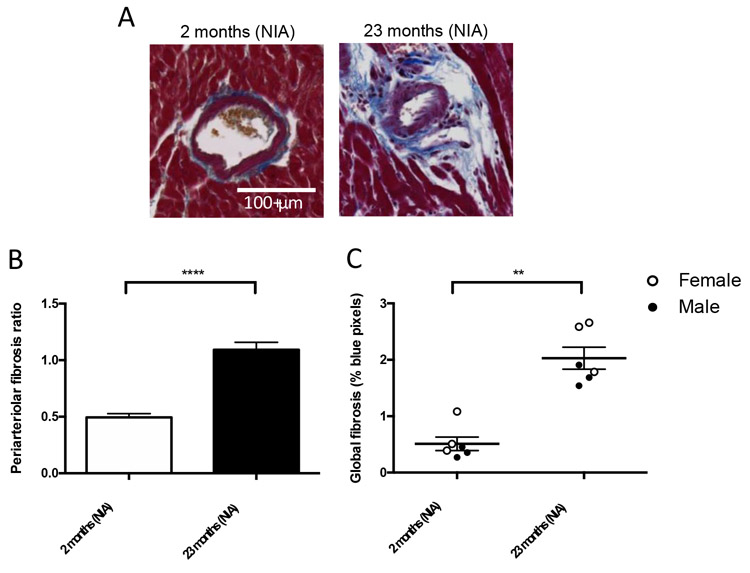
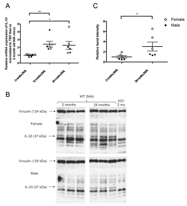
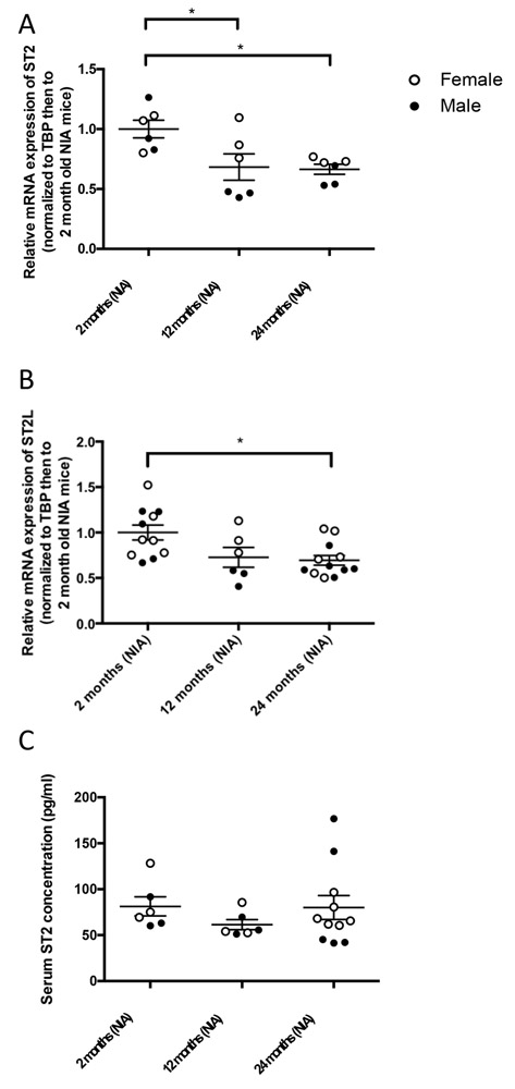
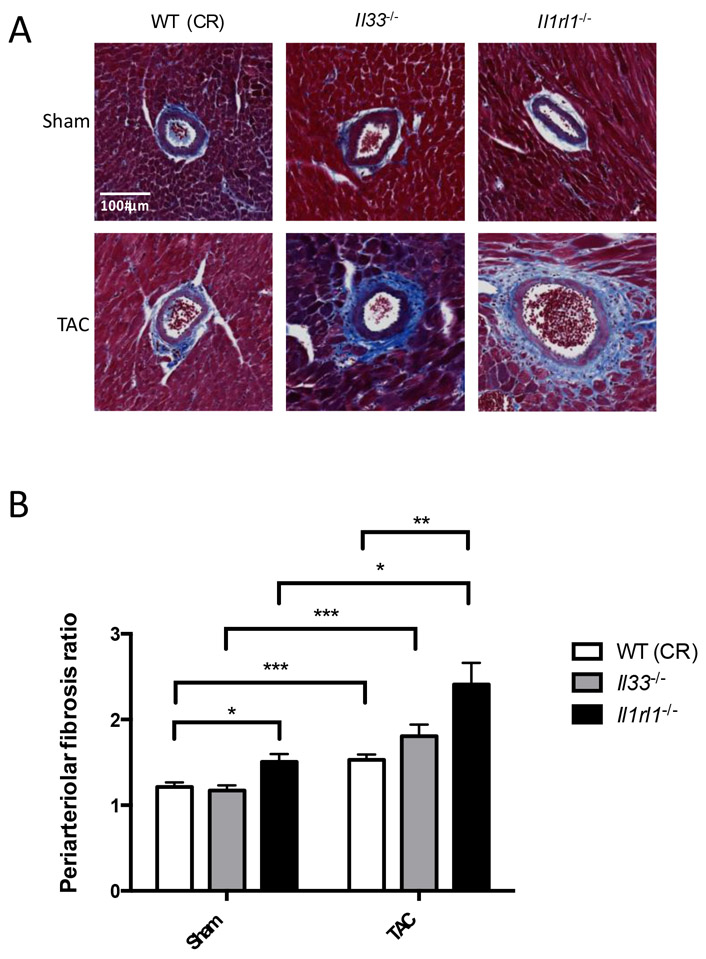
Microvascular dysfunction in the heart and its association with periarteriolar fibrosis may contribute to the diastolic dysfunction seen in heart failure with preserved ejection fraction. Interleukin-33 (IL-33) prevents global myocardial fibrosis in a pressure overloaded left ventricle by acting via its receptor, ST2 (encoded by the gene, Il1rl1); however, whether this cytokine can also modulate periarteriolar fibrosis remains unclear. We utilized two approaches to explore the role of IL-33/ST2 in periarteriolar fibrosis. First, we studied young and old wild type mice to test the hypothesis that IL-33 and ST2 expression change with age. Second, we produced pressure overload in mice deficient in IL-33 or ST2 by transverse aortic constriction (TAC). With age, IL-33 expression increased and ST2 expression decreased. These alterations accompanied increased periarteriolar fibrosis in aged mice. Mice deficient in ST2 but not IL-33 had a significant increase in periarteriolar fibrosis following TAC compared to wild type mice. Thus, loss of ST2 signaling rather than changes in IL-33 expression may contribute to periarteriolar fibrosis during aging or pressure overload, but manipulating this pathway alone may not prevent or reverse fibrosis.
Keywords: Aging; Interleukin-33; Myocardial fibrosis; Periarteriolar fibrosis; Pressure overload; ST2.
Copyright © 2019 Elsevier Ltd. All rights reserved.
Figures
Similar articles
-
Ablation of IL-33 gene exacerbate myocardial remodeling in mice with heart failure induced by mechanical stress.Biochem Pharmacol. 2017 Aug 15;138:73-80. doi: 10.1016/j.bcp.2017.04.022. Epub 2017 Apr 25. Biochem Pharmacol. 2017. PMID: 28450225
-
The Interleukin-33/ST2 Pathway Is Expressed in the Failing Human Heart and Associated with Pro-fibrotic Remodeling of the Myocardium.J Cardiovasc Transl Res. 2018 Feb;11(1):15-21. doi: 10.1007/s12265-017-9775-8. Epub 2017 Dec 28. J Cardiovasc Transl Res. 2018. PMID: 29285671 Free PMC article.
-
ST2/IL-33 signaling in cardiac fibrosis.Int J Biochem Cell Biol. 2019 Nov;116:105619. doi: 10.1016/j.biocel.2019.105619. Epub 2019 Sep 24. Int J Biochem Cell Biol. 2019. PMID: 31561019 Review.
-
IL-33/ST2 Axis in Organ Fibrosis.Front Immunol. 2018 Oct 24;9:2432. doi: 10.3389/fimmu.2018.02432. eCollection 2018. Front Immunol. 2018. PMID: 30405626 Free PMC article. Review.
-
Characterization of a mouse model of obesity-related fibrotic cardiomyopathy that recapitulates features of human heart failure with preserved ejection fraction.Am J Physiol Heart Circ Physiol. 2018 Oct 1;315(4):H934-H949. doi: 10.1152/ajpheart.00238.2018. Epub 2018 Jul 13. Am J Physiol Heart Circ Physiol. 2018. PMID: 30004258 Free PMC article.
Cited by
-
Allograft or Recipient ST2 Deficiency Oppositely Affected Cardiac Allograft Vasculopathy via Differentially Altering Immune Cells Infiltration.Front Immunol. 2021 Mar 18;12:657803. doi: 10.3389/fimmu.2021.657803. eCollection 2021. Front Immunol. 2021. PMID: 33815420 Free PMC article.
-
Full-length IL-33 augments pulmonary fibrosis in an ST2- and Th2-independent, non-transcriptomic fashion.Cell Immunol. 2023 Jan;383:104657. doi: 10.1016/j.cellimm.2022.104657. Epub 2022 Dec 16. Cell Immunol. 2023. PMID: 36603504 Free PMC article.
-
Circulating Soluble ST2 Predicts All-Cause Mortality in Severe Heart Failure Patients with an Implantable Cardioverter Defibrillator.Cardiol Res Pract. 2020 Nov 17;2020:4375651. doi: 10.1155/2020/4375651. eCollection 2020. Cardiol Res Pract. 2020. PMID: 33282418 Free PMC article.
-
Nuclear IL-33 in Fibroblasts Promotes Skin Fibrosis.J Invest Dermatol. 2023 Jul;143(7):1302-1306.e4. doi: 10.1016/j.jid.2022.12.019. Epub 2023 Jan 26. J Invest Dermatol. 2023. PMID: 36708946 Free PMC article. No abstract available.
-
Factors associated with cardiac allograft vasculopathy after heart transplantation.Postepy Kardiol Interwencyjnej. 2022 Sep;18(3):237-245. doi: 10.5114/aic.2022.120370. Epub 2022 Oct 20. Postepy Kardiol Interwencyjnej. 2022. PMID: 36751283 Free PMC article.
References
-
- Dai Z, Aoki T, Fukumoto Y, Shimokawa H, Coronary perivascular fibrosis is associated with impairment of coronary blood flow in patients with non-ischemic heart failure, J Cardiol 60(5) (2012) 416–21. - PubMed
-
- Takayanagi T, Forrester SJ, Kawai T, Obama T, Tsuji T, Elliott KJ, Nuti E, Rossello A, Kwok HF, Scalia R, Rizzo V, Eguchi S, Vascular ADAM17 as a Novel Therapeutic Target in Mediating Cardiovascular Hypertrophy and Perivascular Fibrosis Induced by Angiotensin II, Hypertension 68(4) (2016) 949–955. - PMC - PubMed
Publication types
MeSH terms
Substances
Grants and funding
LinkOut - more resources
Full Text Sources
Medical
Molecular Biology Databases
