. 2019 Feb 5;11(2):181.
doi: 10.3390/cancers11020181.

Cord-Blood-Stem-Cell-Derived Conventional Dendritic Cells Specifically Originate from CD115-Expressing Precursors

Affiliations

Cord-Blood-Stem-Cell-Derived Conventional Dendritic Cells Specifically Originate from CD115-Expressing Precursors

Maud Plantinga et al. Cancers (Basel). .

Abstract

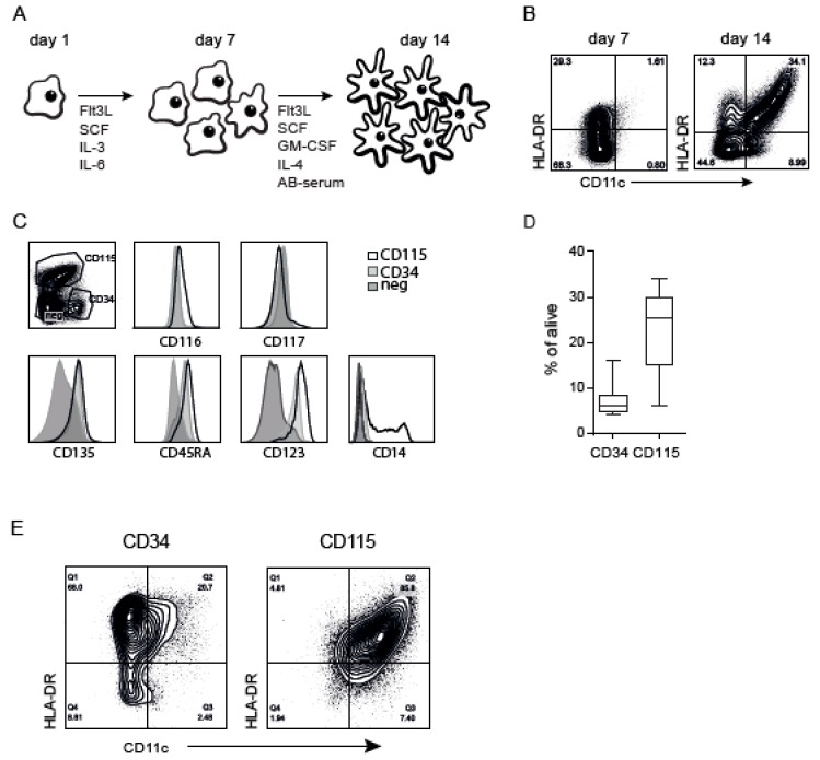
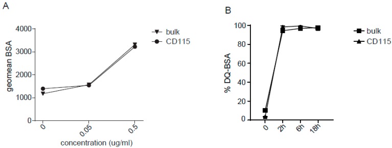
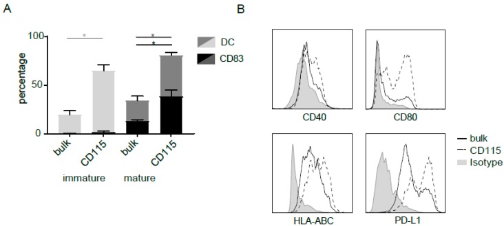
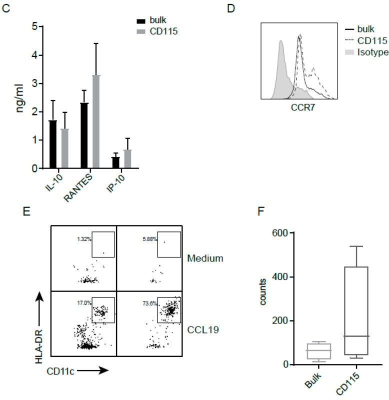
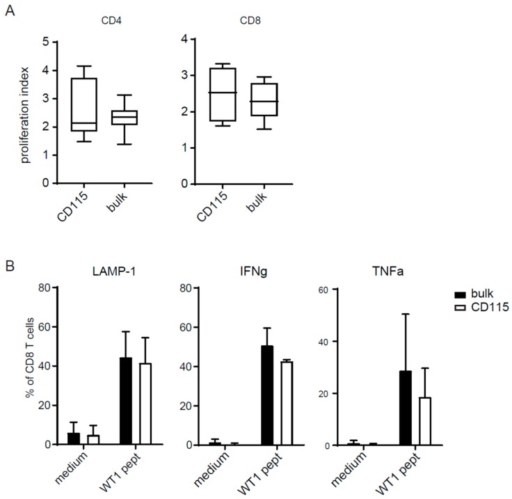
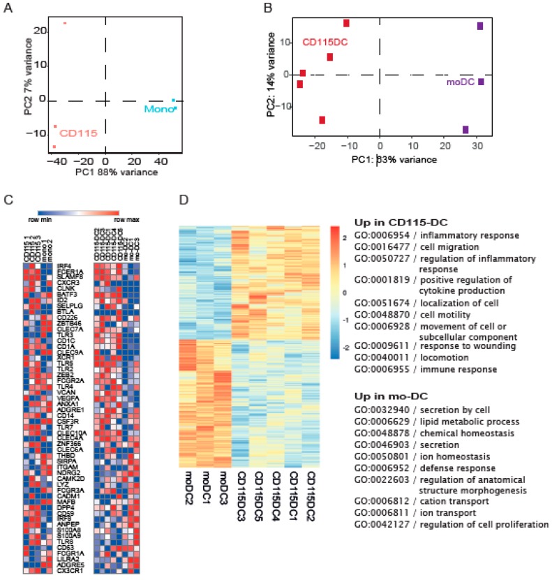
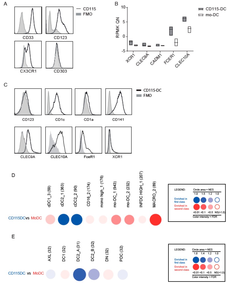
Dendritic cells (DCs) are professional antigen-presenting cells which instruct both the innate and adaptive immune systems. Once mature, they have the capacity to activate and prime naïve T cells for recognition and eradication of pathogens and tumor cells. These characteristics make them excellent candidates for vaccination strategies. Most DC vaccines have been generated from ex vivo culture of monocytes (mo). The use of mo-DCs as vaccines to induce adaptive immunity against cancer has resulted in clinical responses but, overall, treatment success is limited. The application of primary DCs or DCs generated from CD34⁺ stem cells have been suggested to improve clinical efficacy. Cord blood (CB) is a particularly rich source of CD34⁺ stem cells for the generation of DCs, but the dynamics and plasticity of the specific DC lineage development are poorly understood. Using flow sorting of DC progenitors from CB cultures and subsequent RNA sequencing, we found that CB-derived DCs (CB-DCs) exclusively originate from CD115⁺-expressing progenitors. Gene set enrichment analysis displayed an enriched conventional DC profile within the CD115-derived DCs compared with CB mo-DCs. Functional assays demonstrated that these DCs matured and migrated upon good manufacturing practice (GMP)-grade stimulation and possessed a high capacity to activate tumor-antigen-specific T cells. In this study, we developed a culture protocol to generate conventional DCs from CB-derived stem cells in sufficient numbers for vaccination strategies. The discovery of a committed DC precursor in CB-derived stem cell cultures further enables utilization of conventional DC-based vaccines to provide powerful antitumor activity and long-term memory immunity.

Keywords: DC precursor; DC subsets; cancer; cord blood; dendritic cell; vaccine.


Conflict of interest statement

The authors declare no conflicts of interest and the funders had no role in the design of the study; in the collection, analyses, or interpretation of data; in the writing of the manuscript; or in the decision to publish the results.

Figures

Figure 1
Figure 1
Cord blood (CB)-derived dendritic cell (DC) (CB-DC) culture containing different myeloid progenitors. (A) Schematic overview of the culture protocol for the generation of the backbone CB-DC. (B) CD11c and HLA-DR expression using flow cytometry after expansion (day 7) and after differentiation (day 14) within the CB culture. (C) Identification of DC progenitors after the expansion phase based on CD34, CD115. CD116, CD117, CD135, CD45RA, CD123, and CD14 expression compared between CD115+, CD34+, and the remaining population (negative). (D) Percentage of the presence of the progenitors in the CB culture. (E) The expression of CD11c and HLA-DR after differentiating the sorted progenitors in differentiation medium. Data represent at least five independent experiments.
Figure 2
Figure 2
CD115-DCs can take up and process antigen. (A) Uptake of increasing doses of BSA-FITC by DCs from the standard or CD115 culture. The geometric mean of FITC+ signal taken up at 37 °C is shown minus the FITC+ signal detected at 4 °C. (B) The processing of BSA-DQ is shown as a percentage of DQ-BSA gated within the DCs from the standard or CD115 culture at indicated time points. The DQ signal seen at 4 °C is subtracted from the DQ+ cells at 37 °C. Data represent two independent experiments. The experiments were performed at least three times.
Figure 3
Figure 3
Maturation and migration by CD115-DCs. For flow cytometry analysis, live cells were gated with SSC/FSC, followed by doublet exclusion based on FSC-W/FSC-A. (A) Percentages of DCs (HLA-DR+CD11c+) and CD83 at the end of differentiation (day 14) and after an additional 24 h of maturation with cytomix. (B) Expression of CD40, CD80, HLA-ABC, and PD-L1 on DCs (live/single/CD11c+HLA-DR+) from the standard (black line) compared to the CD115 culture (dotted line), with an isotype as control (grey). (C) Cytokines (IL-10, RANTES, and IP-10) produced by standard or CD115 culture after 24 h of maturation. (D) Expression of CCR7 on DCs (live/single/CD11c+HLA-DR+) from the standard (black line) compared to the CD115 culture (dotted line), with an isotype as control (grey). (E) Representative migration assay towards CCL19 or medium as a control with DCs from either the bulk or the CD115 culture. (F) Quantification of migration with four different donors. Count of migrated mature DCs is shown. When indicated, significance was assessed using two-way ANOVA followed by Kruskal–Wallis multiple comparison (* p < 0.05).
Figure 3
Figure 3
Maturation and migration by CD115-DCs. For flow cytometry analysis, live cells were gated with SSC/FSC, followed by doublet exclusion based on FSC-W/FSC-A. (A) Percentages of DCs (HLA-DR+CD11c+) and CD83 at the end of differentiation (day 14) and after an additional 24 h of maturation with cytomix. (B) Expression of CD40, CD80, HLA-ABC, and PD-L1 on DCs (live/single/CD11c+HLA-DR+) from the standard (black line) compared to the CD115 culture (dotted line), with an isotype as control (grey). (C) Cytokines (IL-10, RANTES, and IP-10) produced by standard or CD115 culture after 24 h of maturation. (D) Expression of CCR7 on DCs (live/single/CD11c+HLA-DR+) from the standard (black line) compared to the CD115 culture (dotted line), with an isotype as control (grey). (E) Representative migration assay towards CCL19 or medium as a control with DCs from either the bulk or the CD115 culture. (F) Quantification of migration with four different donors. Count of migrated mature DCs is shown. When indicated, significance was assessed using two-way ANOVA followed by Kruskal–Wallis multiple comparison (* p < 0.05).
Figure 4
Figure 4
(A) T-cell activation was measured in a mixed leukocyte reaction (MLR). Previously isolated CD3 T cells from a different CB donor were thawed and labeled with a cell tracer violet dye. Cells were seeded at 1 × 10⁵ cell/well and stimulated with 2 × 10⁴ cells/well bulk DCs or CD115-DCs for 5 days. Proliferation was measured by FACS and the proliferation index (PI) was calculated using Flowjo. PI is the total number of divisions divided by the number of cells that went into division gated within the CD4 (left) or CD8 (right population). (B) Antigen-specific T-cell activation by sorted CD83+ DCs pulsed o/n with 6 nmol Wilm’s tumor (WT1) peptivator (Miltenyi Biotec, Bergisch Gladbach, Germany) from the CD115 culture compared to the bulk culture. T-cell activation was measured by their intracellular IFNγ and TNFα and extracellular LAMP-1 expression. A represents four different donors and B from two independent experiments.
Figure 5
Figure 5
RNA sequence reveals different genetic profiles within the progenitors and DCs. (AD) RNA-seq data from monocytes, CD115 progenitors, mo-DCs, and CD115-DCs. (A) PCA plot of the CD115 progenitors sorted after 7 days of expansion compared to CD14-isolated monocytes. (B) PCA of CD115-DCs compared to IL-4/GM-CSF-cultured CB-derived mo-DCs. (C) Heatmap generated comparing genes based on prior knowledge on myeloid development and DC subsets using normalized RPMK from RNA-seq data. (D) Heatmap generated from the significantly different genes between mo-DCs and CD115-DCs. Further analysis of the top 500 differentially expressed genes with gene ontology (GO) term analysis using the ToppGene software. The top 10 GO terms of the biological process were upregulated in mo-DCs and CD115-DCs. RNA was isolated from three to five different CB donors. Monocytes and mo-DCs were isolated from two to three CB donors.
Figure 6
Figure 6
Identification of specific CD115 profile. Transcriptome analysis of CD115(-DC) and mo(-DC) was performed by RNA sequencing of sorted precursors or differentiated DCs generated from CB-derived CD34+ stem cells from three to five donors. For FACS analysis, alive cells were gated with SSC/FSC, followed by doublet exclusion based on FSC-W/FSC-A. (A) Protein levels of markers expressed on myeloid precursors studied on CD115 progenitors with a fluorescence minus one (FMO) as negative staining control. (B) Normalized RNA levels of genes expressed by different DC subsets. (C) Protein expression of CD123, CD1a, CD1c, CD141, CLEC9A, CLEC10A, FceR1, and XCR1 within CD115-DCs with FMO control. (D,E) CD115-DCs and mo-DCs were compared for their relative enrichment in publicly available gene sets using Gene set enrichment analysis (GSEA) through the BubbleGUM software. Results are represented as bubbles in a color matching that of the cell subset in which the gene set was enriched. Stronger and more significant enrichments are represented by bigger and darker bubbles, as illustrated in the legend box of the figure. Specifically, the surface area of the bubbles is proportional to the absolute value of the normalized enrichment score (NES). The color intensity of the dots is indicative of the false discovery rate (FDR) statistical value. The gene sets were defined based on those used by Bakdash et al. [22], Balan et al. [12], Segura et al. [23] (D), and Villani et al. [15] (E).

Similar articles

Cited by

References

    1. Feuerstein B., Berger T.G., Maczek C., Roder C., Schreiner D., Hirsch U., Haendle I., Leisgang W., Glaser A., Kuss O., et al. A method for the production of cryopreserved aliquots of antigen-preloaded, mature dendritic cells ready for clinical use. J. Immunol. Methods. 2000;245:15–29. doi: 10.1016/S0022-1759(00)00269-6. - DOI - PubMed
    1. Thurner B., Roder C., Dieckmann D., Heuer M., Kruse M., Glaser A., Keikavoussi P., Kampgen E., Bender A., Schuler G. Generation of large numbers of fully mature and stable dendritic cells from leukapheresis products for clinical application. J. Immunol. Methods. 1999;223:1–15. doi: 10.1016/S0022-1759(98)00208-7. - DOI - PubMed
    1. Schadendorf D., Ugurel S., Schuler-Thurner B., Nestle F.O., Enk A., Brocker E.B., Grabbe S., Rittgen W., Edler L., Sucker A., et al. Dacarbazine (DTIC) versus vaccination with autologous peptide-pulsed dendritic cells (DC) in first-line treatment of patients with metastatic melanoma: A randomized phase III trial of the DC study group of the DeCOG. Ann. Oncol. 2006;17:563–570. doi: 10.1093/annonc/mdj138. - DOI - PubMed
    1. Shinde P., Fernandes S., Melinkeri S., Kale V., Limaye L. Compromised functionality of monocyte-derived dendritic cells in multiple myeloma patients may limit their use in cancer immunotherapy. Sci. Rep. 2018;8:5705. doi: 10.1038/s41598-018-23943-w. - DOI - PMC - PubMed
    1. Prue R.L., Vari F., Radford K.J., Tong H., Hardy M.Y., D’Rozario R., Waterhouse N.J., Rossetti T., Coleman R., Tracey C., et al. A phase I clinical trial of CD1c (BDCA-1)+ dendritic cells pulsed with HLA-A*0201 peptides for immunotherapy of metastatic hormone refractory prostate cancer. J. Immunother. 2015;38:71–76. doi: 10.1097/CJI.0000000000000063. - DOI - PubMed

LinkOut - more resources