Review
. 2019 Feb 5;6(1):14.
doi: 10.3390/bioengineering6010014.

Ear Reconstruction Simulation: From Handcrafting to 3D Printing

Affiliations
Review

Ear Reconstruction Simulation: From Handcrafting to 3D Printing

Elisa Mussi et al. Bioengineering (Basel). .

Abstract

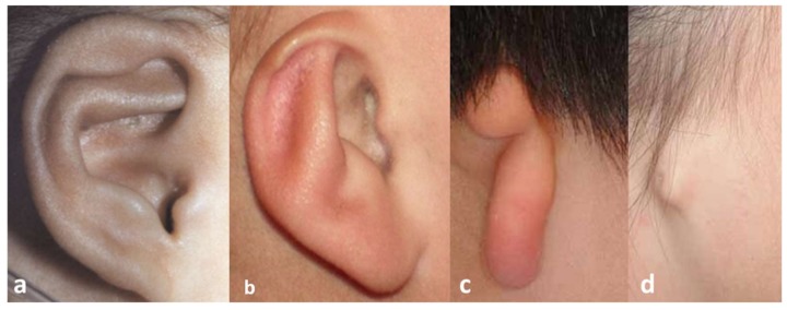
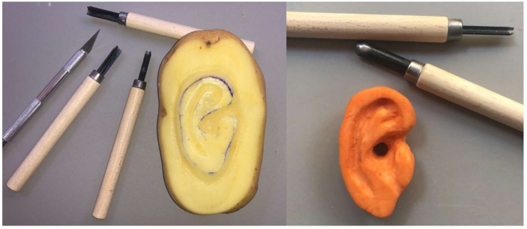
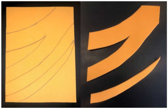
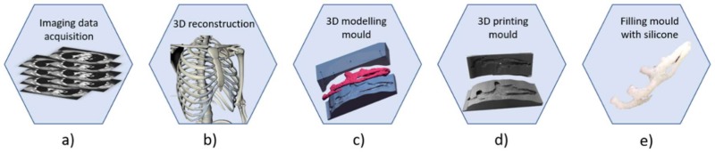
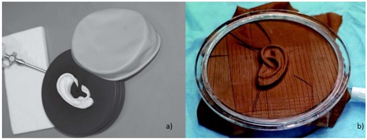
Microtia is a congenital malformation affecting one in 5000 individuals and is characterized by physical deformity or absence of the outer ear. Nowadays, surgical reconstruction with autologous tissue is the most common clinical practice. The procedure requires a high level of manual and artistic techniques of a surgeon in carving and sculpting of harvested costal cartilage of the patient to recreate an auricular framework to insert within a skin pocket obtained at the malformed ear region. The aesthetic outcomes of the surgery are highly dependent on the experience of the surgeon performing the surgery. For this reason, surgeons need simulators to acquire adequate technical skills out of the surgery room without compromising the aesthetic appearance of the patient. The current paper aims to describe and analyze the different materials and methods adopted during the history of autologous ear reconstruction (AER) simulation to train surgeons by practice on geometrically and mechanically accurate physical replicas. Recent advances in 3D modelling software and manufacturing technologies to increase the effectiveness of AER simulators are particularly described to provide more recent outcomes.

Keywords: Computer-Aided Design (CAD); additive manufacturing; autologous ear reconstruction; costal cartilage; image-processing; microtia; silicone rubbers; simulation; training.


Conflict of interest statement

The authors declare that they have no conflict of interest.

Figures

Figure 1
Figure 1
Grade of microtia: (a) grade I, (b) grade II, (c) grade III, (d) grade IV.
Figure 2
Figure 2
Simulation of ear reconstruction with vegetables.
Figure 3
Figure 3
Costal cartilage in plastic eraser.
Figure 4
Figure 4
Workflow to produce an accurate replica of costal cartilage: a) image data acquisition of the thoracic region through with CT scan; b) image processing and 3D reconstruction of ROI (Region Of Interest); c) modelling of mold with 3D modelling software; d) printing of the mold; e) pouring silicone rubbers inside the mold.
Figure 5
Figure 5
2D template of the ear. A translucent X-ray film is placed against the opposite ear and the main’s shape features are hand-traced, creating a 2D projection of the 3D shape.
Figure 6
Figure 6
Workflow to produce three-dimensional ear template: (a) image data acquisition of the healthy ear; (b) image post-processing and digital 3D reconstruction; (c) mirroring of the ear; (d) 3D printing of the model.
Figure 7
Figure 7
Acquisition of the shape of the ear by means of impression material.
Figure 8
Figure 8
Anatomical elements: (a) Pinna (anti-helix, auricular tubercle, scaphoid fossa, and helix), (b) helix, (c) anti-helix.
Figure 9
Figure 9
Process to unroll helix. (a) Import of the STL file in Blender; (b) creation of a bone chain; (c) placement of the bones within the helix; (d) digital straightening of the helix.
Figure 10
Figure 10
Devices to simulate the skin over the ear framework. (a) “Trainer” by Stortz (Tuttlingen, Germany) to simulate the skin over the reconstructed three-dimensional ear framework [28]; (b) Similar trainer developed by Oyama et al. [67].

References

    1. Heike C.L., Luquetti D.V., Hing A.V. Craniofacial Microsomia Overview. University of Washington; Seattle, WA, USA: 1993. - PubMed
    1. Luquetti D.V., Leoncini E., Mastroiacovo P. Microtia-anotia: A global review of prevalence rates. Birth Defects Res. A Clin. Mol. Teratol. 2011;91:813–822. doi: 10.1002/bdra.20836. - DOI - PMC - PubMed
    1. Ross M.T., Cruz R., Hutchinson C., Arnott W.L., Woodruff M.A., Powell S.K. Aesthetic reconstruction of microtia: A review of current techniques and new 3D printing approaches. Virtual Phys. Prototyp. 2018;13:117–130. doi: 10.1080/17452759.2018.1430246. - DOI
    1. Ishimoto S., Ito K., Karino S., Takegoshi H., Kaga K., Yamasoba T. Hearing levels in patients with microtia: Correlation with temporal bone malformation. Laryngoscope. 2007;117:461–465. doi: 10.1097/MLG.0b013e31802ca4d4. - DOI - PubMed
    1. Llano-Rivas I., González-del Angel A., del Castillo V., Reyes R., Carnevale A. Microtia: A clinical and genetic study at the National Institute of Pediatrics in Mexico City. Arch. Med. Res. 1999;30:120–124. doi: 10.1016/S0188-0128(98)00023-2. - DOI - PubMed

LinkOut - more resources