Review
. 2019 Feb 5;7(1):11.
doi: 10.3390/biomedicines7010011.

Ultrastructure in Transthyretin Amyloidosis: From Pathophysiology to Therapeutic Insights

Affiliations
Review

Ultrastructure in Transthyretin Amyloidosis: From Pathophysiology to Therapeutic Insights

Haruki Koike et al. Biomedicines. .

Abstract

Transthyretin (TTR) amyloidosis is caused by systemic deposition of wild-type or variant amyloidogenic TTR (ATTRwt and ATTRv, respectively). ATTRwt amyloidosis has traditionally been termed senile systemic amyloidosis, while ATTRv amyloidosis has been called familial amyloid polyneuropathy. Although ATTRwt amyloidosis has classically been regarded as one of the causes of cardiomyopathy occurring in the elderly population, recent developments in diagnostic techniques have significantly expanded the concept of this disease. For example, this disease is now considered an important cause of carpal tunnel syndrome in the elderly population. The phenotypes of ATTRv amyloidosis also vary depending on the mutation and age of onset. Peripheral neuropathy usually predominates in patients from the conventional endemic foci, while cardiomyopathy or oculoleptomeningeal involvement may also become major problems in other patients. Electron microscopic studies indicate that the direct impact of amyloid fibrils on surrounding tissues leads to organ damage, whereas accumulating evidence suggests that nonfibrillar TTR, such as oligomeric TTR, is toxic, inducing neurodegeneration. Microangiopathy has been suggested to act as an initial lesion, increasing the leakage of circulating TTR. Regarding treatments, the efficacy of liver transplantation has been established for ATTRv amyloidosis patients, particularly patients with early-onset amyloidosis. Recent phase III clinical trials have shown the efficacy of TTR stabilizers, such as tafamidis and diflunisal, for both ATTRwt and ATTRv amyloidosis patients. In addition, a short interfering RNA (siRNA), patisiran, and an antisense oligonucleotide (ASO), inotersen, have been shown to be effective for ATTRv amyloidosis patients. Given their ability to significantly reduce the production of both wild-type and variant TTR in the liver, these gene-silencing drugs seem to be the optimal therapeutic option for ATTR amyloidosis. Hence, the long-term efficacy and tolerability of novel therapies, particularly siRNA and ASO, must be determined to establish an appropriate treatment program.

Keywords: Schwann cell; angiopathy; diflunisal; electron microscopy; oligomers; pathogenesis; pathology; protein misfolding disease; tafamidis; therapy.


Conflict of interest statement

H.K. reports consulting fees and travel fees from Pfizer. M.K. declares no conflicts of interest.

Figures

Figure 1
Figure 1
Representative photographs of cardiac amyloid deposits in early-onset ATTR Val30Met amyloidosis patients from endemic foci (A,B) and late-onset ATTR Val30Met amyloidosis patients from nonendemic areas (C,D) obtained at autopsy. Alkaline Congo red staining. In early-onset patients from endemic foci, the amyloid deposits tend to be highly congophilic (A) and show strong apple-green birefringence (B). In addition, amyloid deposits tend to induce atrophy and degeneration of myocardial cells, particularly in the subendocardial layer, producing a histologic picture of amyloid rings (arrowheads). In late-onset patients from nonendemic areas, the amyloid deposits are generally weakly congophilic (C) and show faint apple-green birefringence (D). Atrophy or degeneration of myocardial cells is not conspicuous in late-onset patients from nonendemic areas compared to early-onset patients from endemic foci. Scale bars = 20 μm.
Figure 2
Figure 2
Representative electron microscopic photographs of amyloid fibrils in early-onset ATTR Val30Met amyloidosis patients from endemic foci (A,C) and late-onset ATTR Val30Met amyloidosis patients from nonendemic areas (B). Cross sections of sural nerve biopsy specimens. Uranyl acetate and lead citrate staining. Amyloid fibrils tend to be long and thick in early-onset patients from endemic foci (A), whereas those in late-onset patients from nonendemic areas are generally short and thin (B). Dotty structures (arrows) are frequently observed among amorphous electron-dense extracellular materials (black arrowheads) (C). Elongated, mature amyloid fibrils are also observed (white arrowheads). Circular structures with a diameter of 50 to 70 nm are collagen fibers. Scale bars = 0.2 μm.
Figure 3
Figure 3
Impact of amyloid fibril formation on neighboring tissues in early-onset ATTR Val30Met amyloidosis patients from endemic foci (A) and late-onset ATTR Val30Met amyloidosis patients from nonendemic areas (B). Cross sections of sural nerve biopsy specimens. Uranyl acetate and lead citrate staining. During the process of amyloid fibril maturation, amyloid fibrils seem to pull surrounding tissues. This traction of neighboring tissues seems to be conspicuous in patients with long and thick amyloid fibrils, such as early-onset Val30Met patients from endemic foci (A). By contrast, the impact of amyloid fibril maturation on neighboring tissues seems to be less in patients with short and fine amyloid fibrils, such as late-onset Val30Met patients from nonendemic areas (B). The stretched basement membrane in (A) is indicated by arrowheads. An unmyelinated fiber in (B) is indicated by an asterisk. Scale bars = 0.5 μm.
Figure 4
Figure 4
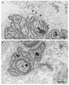
Aggregation of amyloid fibrils and Schwann cells in ATTRv amyloidosis. A cross section of sural nerve biopsy specimen from an early-onset Val30Met patient from an endemic focus. Uranyl acetate and lead citrate staining. Schwann cells associated with unmyelinated fibers that are apposed to amyloid fibrils become atrophic and distorted, whereas myelinated fibers, particularly large myelinated fibers (arrow), tend to be preserved because the apposition of these fibers to amyloid fibril aggregates is usually partial. A high-powered view of representative Schwann cells associated with unmyelinated fibers in the box in (A) is shown in (B). Scale bars = 2 μm (A) and 0.5 μm (B).
Figure 5
Figure 5
Microangiopathy in ATTRv amyloidosis. A cross section of sural nerve biopsy specimen from a late-onset Val30Met patient from a nonendemic area. Uranyl acetate and lead citrate staining. The continuity of endothelial cells of an endoneurial microvessel is lost (arrow), indicating the disruption of the blood–nerve barrier at this site. A high-powered view of the box in (A) is shown in (B). Scale bars = 1 μm (A) and 0.5 μm (B).

References

    1. Pitkänen P., Westermark P., Cornwell G.G., 3rd Senile systemic amyloidosis. Am. J. Pathol. 1984;117:391–399. - PMC - PubMed
    1. Koike H., Misu K., Ikeda S., Ando Y., Nakazato M., Ando E., Yamamoto M., Hattori N., Sobue G., Study Group for Hereditary Neuropathy in Japan Type I (transthyretin Met30) familial amyloid polyneuropathy in Japan: Early- vs. late-onset form. Arch. Neurol. 2002;59:1771–1776. doi: 10.1001/archneur.59.11.1771. - DOI - PubMed
    1. Benson M.D., Kincaid J.C. The molecular biology and clinical features of amyloid neuropathy. Muscle Nerve. 2007;36:411–423. doi: 10.1002/mus.20821. - DOI - PubMed
    1. Planté-Bordeneuve V., Said G. Familial amyloid polyneuropathy. Lancet Neurol. 2011;10:1086–1097. doi: 10.1016/S1474-4422(11)70246-0. - DOI - PubMed
    1. Adams D., Cauquil C., Labeyrie C. Familial amyloid polyneuropathy. Curr. Opin. Neurol. 2017;30:481–489. doi: 10.1097/WCO.0000000000000476. - DOI - PubMed