. 2019 Feb 5;24(3):575.
doi: 10.3390/molecules24030575.

Fluorescent DNA Biosensor for Single-Base Mismatch Detection Assisted by Cationic Comb-Type Copolymer

Affiliations

Fluorescent DNA Biosensor for Single-Base Mismatch Detection Assisted by Cationic Comb-Type Copolymer

Jialun Han et al. Molecules. .

Abstract

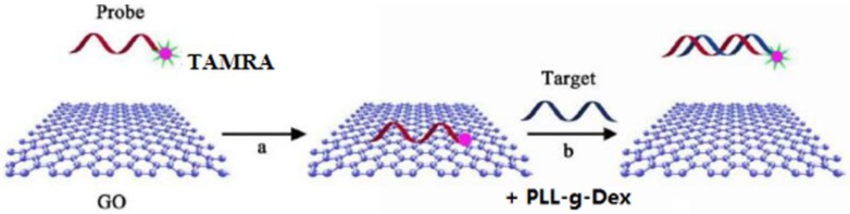
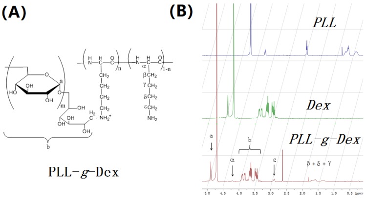
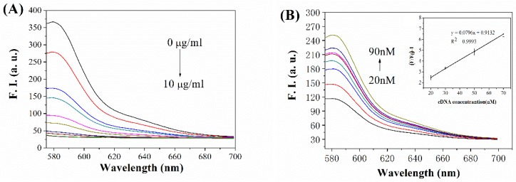
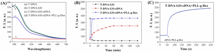
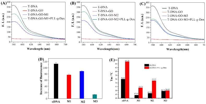
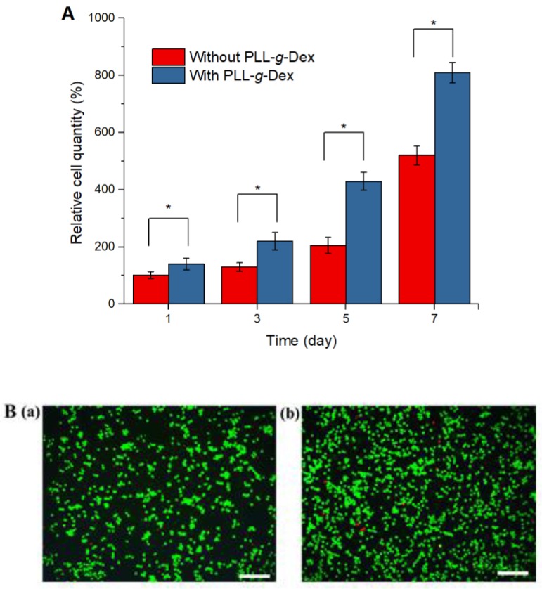
Simple and rapid detection of DNA single base mismatch or point mutation is of great significance for the diagnosis, treatment, and detection of single nucleotide polymorphism (SNP) in genetic diseases. Homogeneous mutation assays with fast hybridization kinetics and amplified discrimination signals facilitate the automatic detection. Herein we report a quick and cost-effective assay for SNP analysis with a fluorescent single-labeled DNA probe. This convenient strategy is based on the efficient quenching effect and the preferential binding of graphene oxide (GO) to ssDNA over dsDNA. Further, a cationic comb-type copolymer (CCC), poly(l-lysine)-graft-dextran (PLL-g-Dex), significantly accelerates DNA hybridization and strand-exchange reaction, amplifying the effective distinction of the kinetic barrier between a perfect matched DNA and a mismatched DNA. Moreover, in vitro experiments indicate that RAW 264.7 cells cultured on PLL-g-Dex exhibits excellent survival and proliferation ability, which makes this mismatch detection strategy highly sensitive and practical.

Keywords: SNP analysis; cationic comb-type copolymer; fluorescent DNA biosensor; graphene oxide; single-base mismatch detection.


Conflict of interest statement

The authors declare no conflict of interest.

Figures

Scheme 1
Scheme 1
Principle for the graphene oxide (GO) based fluorescent DNA biosensor for single-base mismatch detection assisted by cationic comb-type copolymer.
Figure 1
Figure 1
(A) Structural formula of poly(l-lysine)(PLL)-g-Dex. (B) 1H-NMR spectra of PLL, Dex and PLL-g-Dex in D2O. The dextran content of the copolymer was calculated from 1H-NMR signals assigned to PLL (ε-CH2) and dextran (C1-H, a).
Figure 2
Figure 2
(A) The fluorescence intensity of TAMRA labled T-DNA (20 nM) in the presence of various concentrations of GO (0, 2, 3, 4, 5, 6, 7, 8, 9, 10 μg/mL); (B) the fluorescence intensity of T-DNA-GO in the presence of different concentrations of cDNA (20, 30, 40, 50, 60, 70, 80, and 90 nM); (insert) the values of [(F/F0)-1] for assay with the concentration of cDNA.
Figure 3
Figure 3
(A) The fluorescence intensity of T-DNA (20 nM) under the following conditions: (a) T-DNA in the solution; (b) T-DNA + GO (9 μg/mL); (c) T-DNA + GO (9 μg/mL) + cDNA (90 nM); and (d) T-DNA + GO (9 μg/mL) + cDNA (90 nM) + PLL-g-Dex (96 nM). (B) The fluorescence intensity of T-DNA (20 nM) with reactive time under the following conditions: (■) T-DNA + GO (9 μg/mL); (●) T-DNA + GO (9 μg/mL) + cDNA (90 nM); (▲) T-DNA + GO (9 μg/mL) + cDNA (90 nM) + PLL-g-Dex (96 nM). (C) The fluorescence intensity changes in (B) T-DNA + GO (9 μg/mL) + cDNA (90 nM) + PLL-g-Dex (96 nM) within the first 300 s hybridization.
Figure 4
Figure 4
(AC) The fluorescence intensity of target chains with different mismatched bases. (D) The increase of fluorescent intensity at 582 nm in the presence of PLL-g-Dex, compared with the fluorescent intensity at 582 nm without PLL-g-Dex. (E) Tm of different double-strands DNA without (black) or with (red) PLL-g-Dex. [GO (9 μg/mL), cDNA, M1, M2, or M3 (90 nM), PLL-g-Dex (96 nM)].
Figure 5
Figure 5
(A) Proliferation of the RAW 264.7 cells cultured on the surfaces of the PLL-g-Dex or the culture plates (without PLL-g-Dex). * p < 0.05. (B) Viability and morphology of the RAW 264.7 cells cultured on surfaces of (a) the culture plates and (b) PLL-g-Dex for 24 h (green cells are live, and red cells are dead; the scale bar is 200 μm).

Similar articles

Cited by

References

    1. Kashuk C., SenGupta S., Eichler E., Chakravarti A. ViewGene: A graphical tool for polymorphism visualization and characterization. Genome Res. 2002;12:333–338. doi: 10.1101/gr.211202. - DOI - PMC - PubMed
    1. Brennan M.D. High throughput genotyping technologies for pharmacogenomics. Am. J. Pharm. 2001;1:295–302. doi: 10.2165/00129785-200101040-00006. - DOI - PubMed
    1. Kwok P.Y. Methods for genotyping single nucleotide polymorphisms. Annu. Rev. Genom. Hum. Genet. 2001;2:235–258. doi: 10.1146/annurev.genom.2.1.235. - DOI - PubMed
    1. Chicurel M. Faster, better, cheaper genotyping. Nature. 2001;412:580–582. doi: 10.1038/35088146. - DOI - PubMed
    1. Kim W.J., Sato Y., Akaike T., Maruyama A. Cationic comb-type copolymers for DNA analysis. Nat. Mater. 2003;2:815–820. doi: 10.1038/nmat1021. - DOI - PubMed

LinkOut - more resources