

Case Reports
doi: 10.1177/1591019918824796.
Epub 2019 Feb 14.
Vessel wall enhancement of a ruptured intra-nidal aneurysm in a brain arteriovenous malformation
Affiliations
- PMID: 30764685
- PMCID: PMC6547198
- DOI: 10.1177/1591019918824796
Item in Clipboard
Case Reports
Vessel wall enhancement of a ruptured intra-nidal aneurysm in a brain arteriovenous malformation
Interv Neuroradiol.
2019 Jun.
Abstract
Ruptured arteriovenous malformations are a frequently encountered pathology with significant associated morbidity and mortality. Identifying and securing the rupture point is mandatory; however, this can often be difficult. Black blood vessel wall magnetic resonance imaging is a promising technique for identifying ruptured saccular aneurysms and has been used in cases of multiple aneurysms. Here we describe a case of using this imaging technique to identify the rupture point in a ruptured arteriovenous malformation with histopathological correlation.
Keywords: AVM; haemorrhage; histology; vessel wall enhancement.
Figures
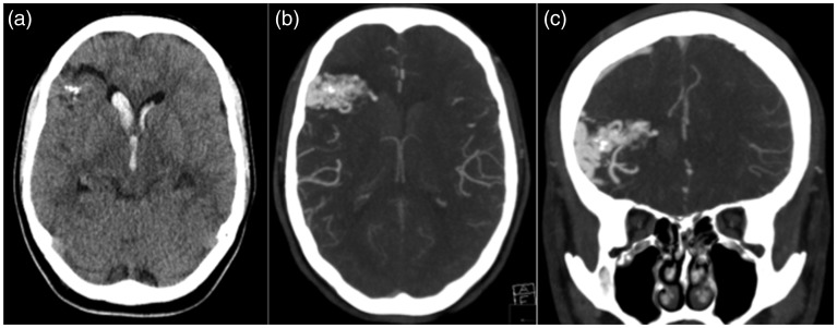
(a) The unenhanced axial computed tomography (CT) scan demonstrates
intraventricular haemorrhage with several foci of calcification within
the right frontal lobe. A CT angiogram demonstrates a right frontal
arteriovenous malformation with an aneurysm directed into the right
frontal horn of the lateral ventricle that was believed to be the source
of the haemorrhage (b and c).
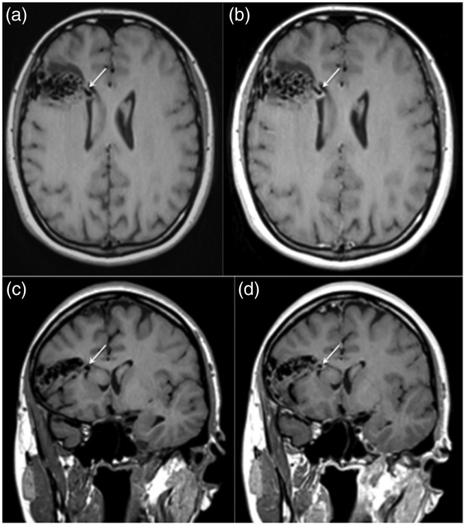
A magnetic resonance image with pre and post-contrast T1-weighted
sequences was performed. The aneurysm demonstrated on the computed
tomography angiogram was clearly visible on the pre-contrast T1-weighted
sequences (a and c, white arrow) with thick, circumferential enhancement
of the aneurysmal wall seen on the post-contrast sequences (b and d,
white arrow). There was no significant enhancement seen elsewhere within
the arteriovenous malformation.
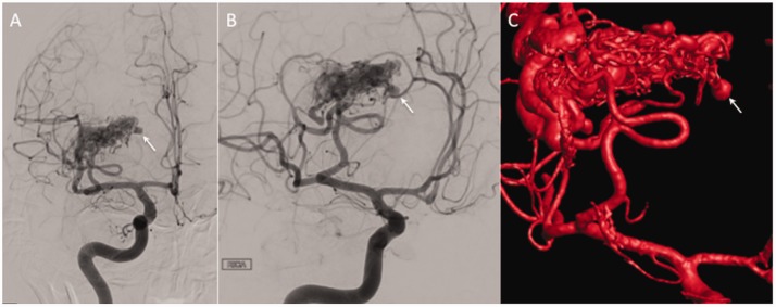
Catheter angiography of the right internal carotid artery demonstrated
supply to the arteriovenous malformation from branches of the middle
cerebral artery (a and b) with a medially directed aneurysm that
correlated with the enhancing aneurysm seen on magnetic resonance
imaging that was clearly appreciated on intra-arterial rotational
angiography (c).
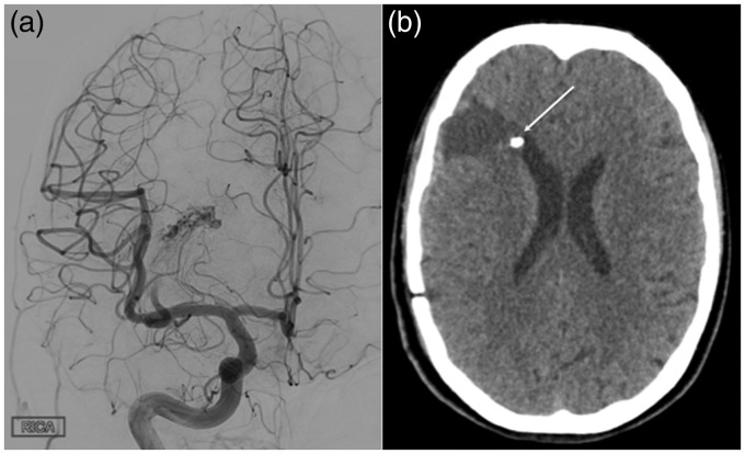
At the end of the embolisation procedure persistent filling of the
superior component of the arteriovenous malformation (AVM) was noted
and further endovascular embolisation was not deemed feasible (a).
The patient underwent resection for the residual AVM. A
postoperative computed tomography scan showed successful resection
of the AVM, with the embolised aneurysm still demonstrable adjacent
to the right frontal horn.
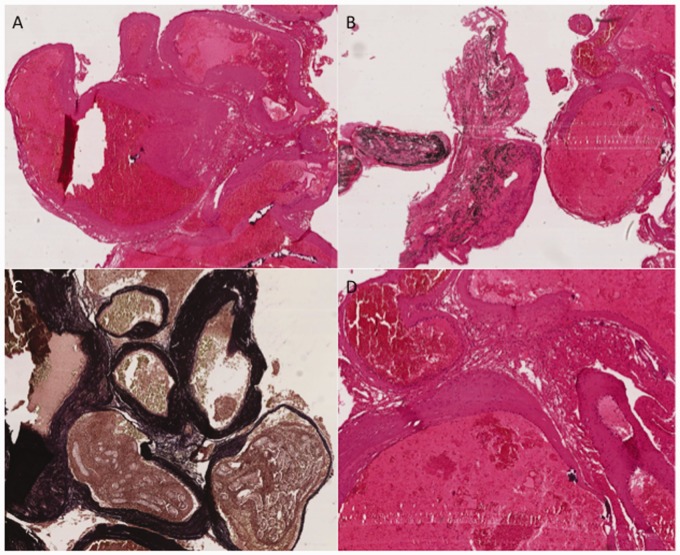
Arteriovenous malformation demonstrating classic histological
appearances of (a) closely associated, sometimes tortuous, ectatic
vascular channels with walls of varying thickness and size.
Occasional vessels contain black-staining embolisation material
(Onyx) (b). Several vessel walls contain prominent internal elastic
laminae, which are demonstrated on Masson trichrome staining as dark
staining within the vessel walls (c). A higher power representative
view demonstrating no histological evidence of mural inflammatory
cell infiltrate within the vessel walls (d).
References
-
- Mast H, Young WL, Koennecke HC, et al. Risk of spontaneous haemorrhage after diagnosis of cerebral arteriovenous malformation. Lancet Lond Engl 1997; 350: 1065–1068. - PubMed
-
- Meisel HJ, Mansmann U, Alvarez H, et al. Cerebral arteriovenous malformations and associated aneurysms: analysis of 305 cases from a series of 662 patients. Neurosurgery 2000; 46: 793–800. discussion 800–802. - PubMed
-
- Hademenos GJ, Massoud TF. Risk of intracranial arteriovenous malformation rupture due to venous drainage impairment. A theoretical analysis. Stroke 1996; 27: 1072–1083. - PubMed
-
- Bhogal P, Uff C, Makalanda HLD. Vessel wall MRI and intracranial aneurysms. J Neurointervent Surg 2016; 8: 1160–1162. - PubMed
Publication types
MeSH terms
Grants and funding
LinkOut - more resources
Full Text Sources
Medical
