Review
. 2018 Dec;243(17-18):1245-1255.
doi: 10.1177/1535370218820287. Epub 2019 Jan 6.

The great escape: How metastases of melanoma, and other carcinomas, avoid elimination

Affiliations
Review

The great escape: How metastases of melanoma, and other carcinomas, avoid elimination

Alan Wells et al. Exp Biol Med (Maywood). 2018 Dec.

Abstract

Cancers kill mainly because metastatic disease is resistant to systemic therapies. It was hoped that newer targeted and immunomodulatory interventions could overcome these issues. However, recent findings point to a generalized resistance to elimination imparted by both cancer-intrinsic and -extrinsic changes to provide survival advantages to the disseminated tumor cells. Here, we present a novel conceptual framework for the microenvironmental inputs and changes that contribute to this generalized therapeutic resistance. In addition we address the issues of experimental systems in terms of studying this phenomenon with their advantages and limitations. This is meant to spur studies into this critical aspect of tumor progression that directly leads to cancer mortality.

Keywords: Liver metastasis; hepatic niche; metastatic models; microphysiological; tumor microenvironment.


Figures

Figure 1.
Figure 1.
Metastatic cascade to the liver. Schematic of metastasis with postulated phenotypes that confer resistance or sensitivity to therapies. Disseminating carcinoma cells must acquire mesenchymal-like migratory properties to escape the primary locale (the cEMT). Transit through vascular conduits to sites of metastasis are fraught with challenges with most tumor cells not surviving, but are sufficiently transient to not represent a target for therapy. At the metastatic site, cells must survive apoptotic cytokines initially and chemotherapy later; this is accomplished by a reversion to a more epithelial phenotype and expression of E-cadherin (cMErT) that in turn provides for the chemoresistance and immune silence (PD-L1-negative), driven at least in part by host organ extracellular vesicles (green bubbles). Cells emerge from dormancy due to inflammatory stimuli (lightning bolts) to form aggressive, lethal metastatic nodules with re-acquisition of mesenchymal-like behaviors and cause the organ to secrete factors with secondary metastases also ensuing (adapted from Clark et al). (A color version of this figure is available in the online journal.)
Figure 2.
Figure 2.
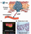
Mechanisms of therapeutic resistance in the primary tumor. (i) Escape from the primary tumor site is promoted by the tumor microenvironment being altered with upregulation of motility-promoting matricellular and matrix-associated proteins (e.g. TNC). TNC is at the invasive front as demonstrated in (ii) an invading melanoma cell lines and (iii) a patient melanoma specimen (adapted from Grahovac et al). However, as these cells are highly active, these protections from death are minimal in the pre-disseminated site.
Figure 3.
Figure 3.
Mechanisms of therapeutic resistance in the micrometastases. (i) Micrometastases escape therapy by upregulation of survival and downregulation of target molecules. (ii) Tumor cells expressing high levels of the E-cadherin in the liver are more resistant to chemotherapy than those expressing low levels (adapted from Ma et al). (iii) An inverse relationship between E-cadherin (chemoresistant) and PD-L1 proteins (here shown in experimental metastasis models in the mouse, unpublished data) would obviate immunotherapy targeting. (iv) EVs from the organ tissue of the metastatic site, particularly the non-parenchymal immune and stromal cells, drive the conversion cMErT in colonizing disseminated cells (adapted from Dioufa et al). (A color version of this figure is available in the online journal.)
Figure 4.
Figure 4.
Mechanisms of therapeutic resistance in the macrometastases. (i) Mechanisms of therapeutic resistance persists through a second cEMT and subsequent overt growth. (ii) Therapeutic resistance of tumor cells is maintained via the production of the same matricellular and matrix-associated proteins (e.g. TNC) produced during the first cEMT event for invasion with this now encasing the entire metastasis. (iii) E-cadherin expression inversely correlates with tumor size, with the loss of E-cadherin tracking with emergence as a clinically evident metastatic nodule, opening the way for greater therapeutic sensitivity (adapted from Chao et al). (A color version of this figure is available in the online journal.)

References

    1. Mieog JS, vanderHage JA, vandeVelde CJ. Neoadjuvant chemotherapy for operable breast cancer. Br J Surg 2007; 94:1189–200 - PubMed
    1. Redden MH, Fuhrman GM. Neoadjuvant chemotherapy in the treatment of breast cancer. Surg Clin North Am 2013; 93:493–9 - PubMed
    1. Mougalian SS, Hernandez M, Lei X, Lynch S, Kuerer HM, Symmans W, Theriault RL, Fornage BD, Hsu L, Buchholz TA, Sahin AA, Hunt KK, Yang WT, Hortobagyi GN, Valero V. Ten-year outcomes of patients with breast cancer with cytologically confirmed axillary lymph node metastases and pathologic complete response after primary systemic chemotherapy. JAMA Oncol 2016; 2:508–16 - PMC - PubMed
    1. Fidler IJ. The pathogenesis of cancer metastasis: the 'seed and soil' hypothesis revisited. Nat Rev Cancer 2003; 3:453–8 - PubMed
    1. Clark AM, Ma B, Taylor DL, Griffith L, Wells A. Liver metastases: microenvironments and ex vivo models. Exp Biol Med 2016; 241:1639–52 - PMC - PubMed

Publication types