. 2019 Mar 7;176(6):1490-1501.e12.
doi: 10.1016/j.cell.2019.02.002. Epub 2019 Feb 11.

Mechanism of Cross-talk between H2B Ubiquitination and H3 Methylation by Dot1L

Affiliations

Mechanism of Cross-talk between H2B Ubiquitination and H3 Methylation by Dot1L

Evan J Worden et al. Cell. .

Abstract

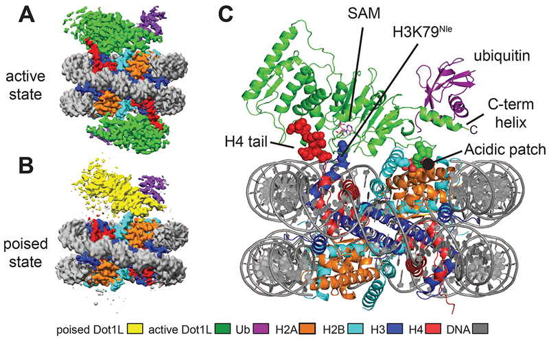
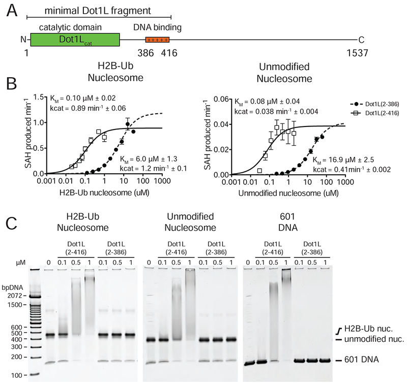
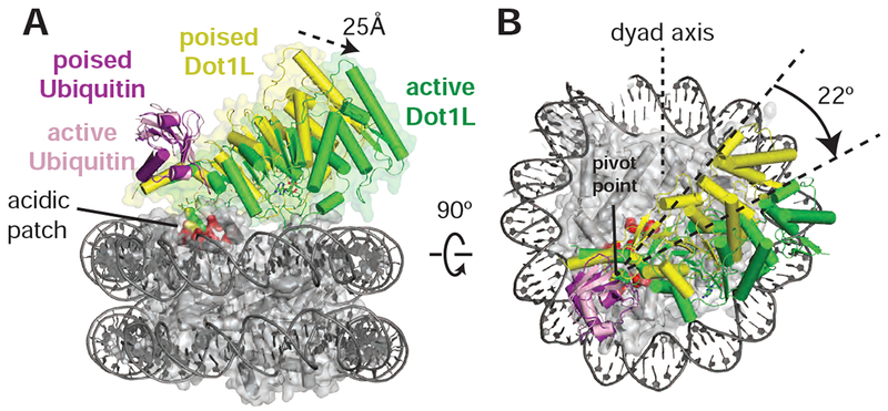
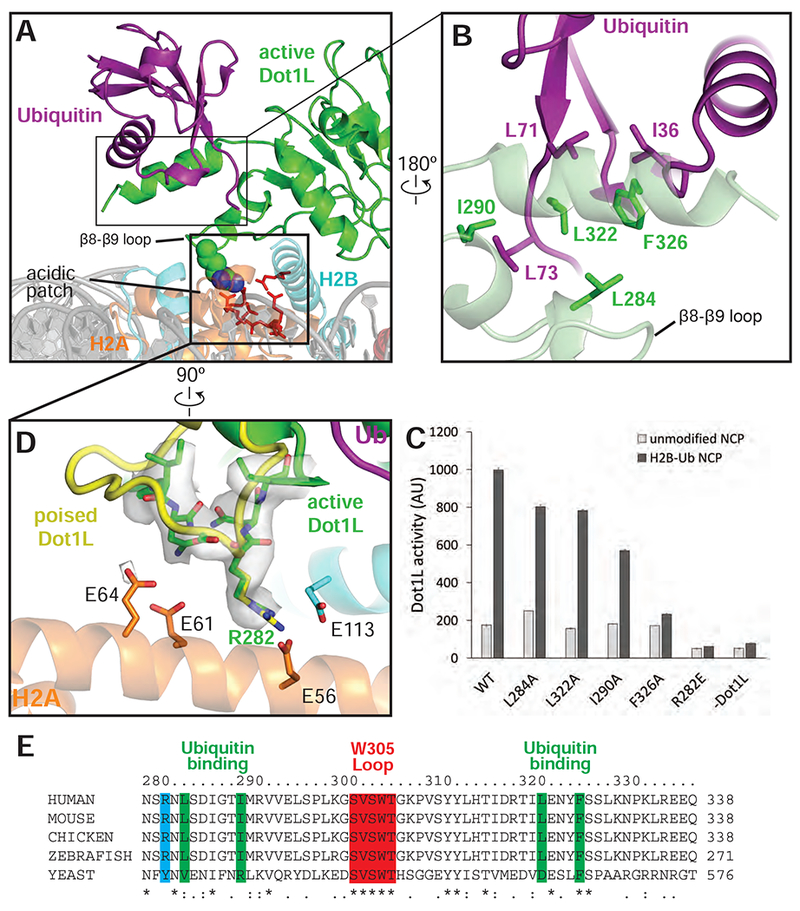
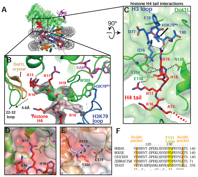
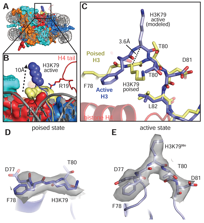
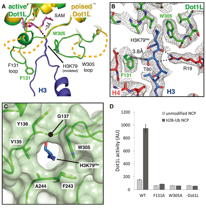
Methylation of histone H3 K79 by Dot1L is a hallmark of actively transcribed genes that depends on monoubiquitination of H2B K120 (H2B-Ub) and is an example of histone modification cross-talk that is conserved from yeast to humans. We report here cryo-EM structures of Dot1L bound to ubiquitinated nucleosome that show how H2B-Ub stimulates Dot1L activity and reveal a role for the histone H4 tail in positioning Dot1L. We find that contacts mediated by Dot1L and the H4 tail induce a conformational change in the globular core of histone H3 that reorients K79 from an inaccessible position, thus enabling this side chain to insert into the active site in a position primed for catalysis. Our study provides a comprehensive mechanism of cross-talk between histone ubiquitination and methylation and reveals structural plasticity in histones that makes it possible for histone-modifying enzymes to access residues within the nucleosome core.

Keywords: Dot1L; chromatin; cryo-EM; histones; methylation; nucleosome; structural biology; ubiquitin.


Conflict of interest statement

Competing Interests

C.W. is a member of the scientific advisory board of ThermoFisher Scientific.

Figures

Figure 1:
Figure 1:. Structures Dot1L bound to H2B-ubiquitin nucleosome
A, Cryo-EM reconstruction of the 2-to-1 active state complex. B, Cryo-EM reconstruction of the poised state complex. C, Atomic model of the active state complex between Dot1L and the H2B-Ub nucleosome. The H4 tail, H3K79Nle and the acidic patch are depicted as spheres and the SAM cofactor is depicted in stick representation. See also Figures S1–S4.
Figure 2 :
Figure 2 :. Analysis of Dot1L binding and activity.
A, Schematic of the Dot1L domain architecture. B Michaelis Menten titrations of two variants of Dot1L. Left, titration with H2B-Ub nucleosome. Right, titration with unmodified Nucleosome. Error bars correspond to the standard error of three replicate measurements. The kcat and Km of the fitted data are reported in the graph field. The reported errors of the fitted Km and kcat correspond to the standard error. C Electrophoretic Mobility Shift Assay of Dot1L variants binding to different nucleosome substrates and to free DNA.
Figure 3:
Figure 3:. Dot1L transitions between the Poised and Active States
Dot1L movements from the poised state to the active state shown as from the side , A, and top, B. Poised state (yellow) and active state (green) Dot1L are depicted as cylinders and the histone octamer is shown as a transparent gray surface. H2A/H2B acidic patch residues are shown as red spheres. Arrows indicate movements made by Dot1L in the switch to the active state. See also Figure S3 and S5.
Figure 4:
Figure 4:. Dot1L interactions with Ubiquitin and the acidic patch
A, Overview of the interaction between Dot1L, Ubiquitin and the nucleosome in the active state structure. Residues important for Dot1L interaction with the nucleosome acidic patch residues are depicted as red sticks. B, Detailed view of the contacts made between Dot1L and ubiquitin. Dot1L is depicted as a semi-transparent green cartoon. Important residues at the Dot1L-Ubiquitin interface are shown as sticks. C, Endpoint H3K79 methylation activity assays using Dot1L mutants with either unmodified or H2B-Ub nucleosomes. Error bars correspond to the standard deviation of 3 replicate experiments. D, Detailed view of interactions between Dot1L and the H2B/H2A acidic patch. Residues at the interface are depicted as sticks and the EM density for Dot1L in the active state is shown as a semi-transparent gray surface. A superimposed poised state Dot1L is depicted in yellow. E, Multiple Sequence alignment of Dot1L from different species. The alignment was performed with Clustal Omega (Sievers et al., 2011). See also Figures S3, S5 and Table S1.
Figure 5:
Figure 5:. H4 tail interactions with Dot1L
A, Overview of the active Dot1L structure. Dot1L is shown as a transparent green surface, ubiquitin as purple ribbon, H4 tail in red spheres. B, H4 tail (red) interaction with the Dot1L binding groove. EM density for the H4 tail is shown as a semi-transparent gray surface. The 22-32 loop from the crystal structure of Dot1L alone (PDB ID 1NW3) is colored tan. C, H4 tail (red) interactions between Dot1L (green) and the H3K79 loop (blue). The surface of Dot1L is shown in semi-transparent green. Potential hydrogen bonding or van der Waals interactions are shown as black dashed lines. Red dashed lines illustrate the direction of the H4 mainchain in the Dot1L binding groove. D, Modeled position of H4 R17 binding in the Dot1L acidic pocket. Conserved residues in the binding pocket are depicted as sticks and the surface of Dot1L is shown and colored according the electrostatic potential. E, Close-up view yeast Dot1p (PDB ID 1U2Z) showing arginine from a neighboring Dot1p molecule in the crystal bound in the conserved acidic pocket. Electrostatic surface potential shown as in D. Electrostatic potential in D, E, was calculated using the APBS tool (Baker et al., 2001). F, Multiple Sequence alignment of Dot1L from different species. The alignment was performed with Clustal Omega (Sievers et al., 2011). See also Figure S6 and Table S1.
Figure 6:
Figure 6:. Conformational change in histone H3 reorients K79
A, Superimposition of the active state and poised state nucleosomes. The active state histone octamer is depicted in surface representation and colored as in Figure 1. The H3K79 loops from the active state (blue) and poised state (yellow) structures are shown as cartoons and H3K79 is shown as spheres. B, Close up view of H3K79 in the active state (blue spheres) and poised state (yellow spheres). The H3K79 sidechain moves ~10 Å (measured from ε-amino groups) from the poised state to the active state. C, Superimposition of the histone H3K79 loop from the active (blue) and poised state (yellow) structures showing the conformational change that occurs in the transition from the poised state to the active state. In the transition from the poised state to the active state the H3K79 backbone moves up by 3.6 Å (measured from the Cα carbons of H3K79) and rotates by an angle of ~90°. D, EM density (gray surface) for the H3K79 loop in the poised state. E, EM density (gray surface) for the H3K79 loop in the active state. See also Figure S7 and Table S1.
Figure 7:
Figure 7:. Formation of the Dot1L active site enclosure
A, Superimposition of active state (green) and poised state (yellow) Dot1L. The H3K79 loop from the active state structure is shown as a blue cartoon and the modeled H3K79 sidechain is shown in stick representation. The disordered F131 and W305 loops from the poised state structure are depicted as yellow dashed lines. The ε- amino group of lysine comes within 3 Å of the SAM methyl donor. B, Close up view of the Dot1L active site enclosure with H3 (blue), Dot1L (green) and H4 (red) shown in stick representation. Sharpened experimental EM density is shown as a gray mesh. A van der Waals contact between Dot1L F131 and H3 T80 is shown as thick a dashed black line and the hydrogen bonds between R19 and the H3K79 loop are shown as thin black dashed lines. C, Formation of the Dot1L H3K79 lysine binding channel. Dot1L is depicted as a green cartoon surrounded by a semi-transparent green surface. H3K79Nle is shown as blue sticks. D, Endpoint H3K79 methylation activity assays using Dot1L mutants with either unmodified or H2B-Ub nucleosomes. Error bars correspond to the standard deviation of 3 replicates. See also Figure S7 and Table S1.

References

    1. Adams PD, Afonine PV, Bunkóczi G, Chen VB, Davis IW, Echols N, Headd JJ, Hung LW, Kapral GJ, Grosse-Kunstleve RW, et al. (2010). PHENIX: A comprehensive Python-based system for macromolecular structure solution. Acta Crystallographica Section D: Biological Crystallography 66, 213–221. - PMC - PubMed
    1. Afonine PV, Poon BK, Read RJ, Sobolev OV, Terwilliger TC, Urzhumtsev A, and Adams PD (2018). Real-space refinement in PHENIX for cryo-EM and crystallography. Acta Crystallogr D Struct Biol 74, 531–544. - PMC - PubMed
    1. Altaf M, Utley RT, Lacoste N, Tan S, Briggs SD, and Côté J (2007). Interplay of Chromatin Modifiers on a Short Basic Patch of Histone H4 Tail Defines the Boundary of Telomeric Heterochromatin. Molecular Cell 28, 1002–1014. - PMC - PubMed
    1. Armache KJ, Garlick JD, Canzio D, Narlikar GJ, and Kingston RE (2011). Structural basis of silencing: Sir3 BAH domain in complex with a nucleosome at 3.0 A resolution. Science 334, 977–982. - PMC - PubMed
    1. Baker NA, Sept D, Joseph S, Holst MJ, and McCammon JA (2001). Electrostatics of nanosystems: application to microtubules and the ribosome. Proc Natl Acad Sci U S A 98, 10037–10041. - PMC - PubMed

Publication types

MeSH terms

LinkOut - more resources