. 2019 Apr;15(4):543-552.
doi: 10.1016/j.jalz.2018.12.003. Epub 2019 Feb 11.

Clinical and cortical decline in the aphasic variant of Alzheimer's disease

Affiliations

Clinical and cortical decline in the aphasic variant of Alzheimer's disease

Emily Joy Rogalski et al. Alzheimers Dement. 2019 Apr.

Abstract

Introduction: Primary progressive aphasia (PPA) displays variable progression trajectories that require further elucidation.

Methods: Longitudinal quantitation of atrophy and language over 12 months was completed for PPA patients with and without positive amyloid PET (PPAAβ+ and PPAAβ-), an imaging biomarker of underlying Alzheimer's disease.

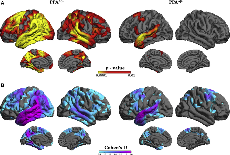
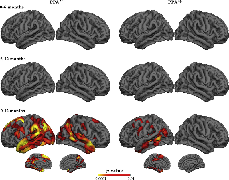
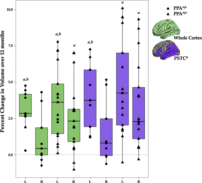
Results: Over 12 months, both PPA groups showed significantly greater cortical atrophy rates in the left versus right hemisphere, with a more widespread pattern in PPAAβ+. The PPAAβ+ group also showed greater decline in performance on most language tasks. There was no obligatory relationship between the logopenic PPA variant and amyloid status. Effect sizes from quantitative MRI data were more robust than neuropsychological metrics.

Discussion: Preferential language network neurodegeneration is present in PPA irrespective of amyloid status. Clinical and anatomical progression appears to differ for PPA due to Alzheimer's disease versus non-Alzheimer's disease neuropathology, a distinction that may help to inform prognosis and the design of intervention trials.

Keywords: Amyloid PET; Biomarker; FreeSurfer; Frontotemporal dementia; Frontotemporal lobar degeneration; Neuropsychology; Primary progressive aphasia; Progression; Volumetric MRI.


Figures

Fig. 1.
Fig. 1.
Characterization of cortical atrophy at initial visit for the PPAAβ+ and PPAAβ− groups. (A) Vertex-based cortical atrophy patterns by amyloid status at the initial visit. Left: Red/yellow indicates significant cortical thinning patterns for the PPAAβ+ participants relative to controls. Right: Red/yellow indicates significant cortical thinning patterns for the PPAAβ− participants relative to controls. The false discovery rate was set at 0.05 for each comparison. (B) Cohn’s d effect-size maps by amyloid status at initial visit based on standard deviation for the PPAAβ+ (left) and PPAAβ− (right) participants. Abbreviations: PPA, primary progressive aphasia.
Fig. 2.
Fig. 2.
Vertex-based percent change in cortical thickness by amyloid status. Red/yellow indicates significant cortical thinning patterns. The false discovery rate was set at 0.05 for each comparison. Abbreviation: PPA, primary progressive aphasia.
Fig. 3.
Fig. 3.
Percent change over 12 months by hemisphere, amyloid status, and ROI. aPercent change in volume was significantly different from zero (P <.0125). bPercent change in volume was significantly greater in the left hemisphere than the right hemisphere (P <.0071). The middle, upper, and lower horizontal lines in the box plot represent the median, 75th, and 25th percentiles, respectively. The end of the vertical extensions above and below the box represent the extreme value that is at most 1.5 times the interquartile range above the 75th percentile or below the 25th percentile. Abbreviations: PPA, primary progressive aphasia; PSTCR, revised language network ROI defined for the perisylvian cortex; ROI, region of interest.

References

    1. Mesulam MM. Primary Progressive Aphasia: A Language-Based Dementia. N Engl J Med 2003;349:1535–42. - PubMed
    1. Gorno-Tempini ML, Dronkers NF, Rankin KP, Ogar JM, Phengrasamy L, Rosen HJ, et al. Cognition and anatomy in three variants of primary progressive aphasia. Ann Neurol 2004;55:335–46. - PMC - PubMed
    1. Mesulam M, Wieneke C, Rogalski E, Cobia D, Thompson C, Weintraub S. Quantitative template for subtyping primary progressive aphasia. Arch Neurol 2009;66:1545–51. - PMC - PubMed
    1. Rogalski E, Sridhar J, Rader B, Martersteck A, Chen K, Cobia D, et al. Aphasic variant of Alzheimer disease: Clinical, anatomic, and genetic features. Neurology 2016;87:1337–43. - PMC - PubMed
    1. Mesulam MM, Weintraub S, Rogalski EJ, Wieneke C, Geula C, Bigio EH. Asymmetry and heterogeneity of Alzheimer’s and frontotemporal pathology in primary progressive aphasia. Brain 2014; 137:1176–92. - PMC - PubMed

Publication types