Review
. 2019 Nov 1;11(11):a032417.
doi: 10.1101/cshperspect.a032417.

Structural Insights into Nuclear pre-mRNA Splicing in Higher Eukaryotes

Affiliations
Review

Structural Insights into Nuclear pre-mRNA Splicing in Higher Eukaryotes

Berthold Kastner et al. Cold Spring Harb Perspect Biol. .

Abstract

The spliceosome is a highly complex, dynamic ribonucleoprotein molecular machine that undergoes numerous structural and compositional rearrangements that lead to the formation of its active site. Recent advances in cyroelectron microscopy (cryo-EM) have provided a plethora of near-atomic structural information about the inner workings of the spliceosome. Aided by previous biochemical, structural, and functional studies, cryo-EM has confirmed or provided a structural basis for most of the prevailing models of spliceosome function, but at the same time allowed novel insights into splicing catalysis and the intriguing dynamics of the spliceosome. The mechanism of pre-mRNA splicing is highly conserved between humans and yeast, but the compositional dynamics and ribonucleoprotein (RNP) remodeling of the human spliceosome are more complex. Here, we summarize recent advances in our understanding of the molecular architecture of the human spliceosome, highlighting differences between the human and yeast splicing machineries.


Figures

Figure 1.
Figure 1.
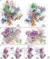
Assembly pathway, RNA network, and compositional dynamics of the spliceosome. (A) Conserved sequences at the 5′ splice site (SS), branch site (BS), and 3′SS of U2-type (major) pre-mRNA introns in metazoans and the yeast Saccharomyces cerevisiae. (B) Schematic of the two-step splicing reaction. p, phosphates at the 5′ and 3′ splice sites, which are conserved in the splicing products. (C) Assembly, catalytic activation, and disassembly pathway of the spliceosome. For simplicity, the ordered interactions of the U1, U2, U4/U6, and U5 small nuclear ribonucleoproteins (snRNPs), but not non-snRNP spliceosomal proteins, are shown. Helicases required for splicing in both yeast and humans are indicated and include the Ski2-like helicase BBR2, the DEAD-box helicases UAP56, PRP5, and PRP28, and the DEAH-box helicases PRP2, PRP16, PRP22, and PRP43. (D) Dynamic network of RNA–RNA interactions in the spliceosomal B and Bact complexes. (E) 3D structure of the catalytic RNA network (in the human C* complex), showing the coordination of the catalytic magnesium ions M1 and M2. (F) Dynamic exchange of spliceosomal proteins during splicing. Proteins present in pre-B, B, Bact, C, or C* human spliceosomal complexes are indicated by a square, in which blue denotes highly abundant and gray moderately abundant proteins. Serine-arginine (SR) dipeptide-rich proteins and heterogeneous nuclear ribonucleoproteins (hnRNPs), as well as those present in very low amounts, are not shown. The table summarizes the proteomes of various, recently purified human spliceosomal complexes (Agafonov et al. 2011; De et al. 2015; Boesler et al. 2016; Bertram et al. 2017b), as well as our unpublished work. *Prp38 and Snu23 are associated with the tri-snRNP in S. cerevisiae. (G) Differential pre-organization of yeast nineteen complex (NTC) proteins in humans. (H) Alternative secondary structures of the U2 small nuclear RNA (snRNA).
Figure 2.
Figure 2.
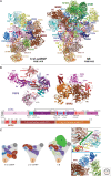
Structure of the human tri-snRNP and spliceosomal B complex. (A) Overview of the structure of the human U4/U6.U5 tri-snRNP and B complex. (B) Schematic of the domain organization of human PRP8 and BRR2. PRP8 contains an amino-terminal domain (NTD), helical bundle (HB), reverse transcriptase-like (RT) domain, and thumb/X domain, followed by a Linker region and an endonuclease-like (En) domain, plus carboxy (C)-terminal RNase-H-like (RH) and Jab1/MPN domains. The RT, En, RH, and Jab1/MPN domains are deconstructed enzymes that do not show enzymatic activities. Within the Linker, several distinct structural elements play important roles within the spliceosome including the switch-loop and the α-finger. The 3D structure of PRP8 shown is from the hB complex, and that of BRR2 (except for its PWI domain) is from hBact. (C) Cartoon of the organization of selected proteins in the human and yeast tri-snRNPs, and human B complex. (D) Upper panel: FBP21’s zinc finger contacts the U6/intron helix, whereas its α-helix keeps U4/U6 stem I, via PPIH (shown as a black outline), at a distance from Brr2NC. Lower panel: SMU1 and RED bridge BRR2 and SF3B3.
Figure 3.
Figure 3.
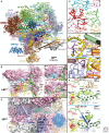
Molecular organization of RNA−protein interactions involving the catalytic RNA network, 5′SS, and 5′ exon in the human Bact complex. (A) Overview of the structural organization of the human Bact complex. The positions of PPIases are circled in black. (B) Accommodation of the catalytic RNA network that is first formed in Bact by the PRP8 amino-terminal domain (NTD) and helical bundle (HB) domains and α-finger of its Linker domain. Part of RNF113A (Cwc24) and SF3A2 (Prp11) are inserted into the gap between the RT/En domain and the catalytic RNA network and these proteins are replaced by the U2/BS helix in the C complex (see insets). (C) Close-up of interactions between the 5′SS nucleotides G+1 and U+2 and residues of RNF113A, and between SF3A2 and G+1 and G−1. (D) PRP19/CDC5L complex proteins and related proteins directly contact primarily flipped-out U6 nucleotides in the catalytic RNA core. For example, the base of U6-C60 in the lower stem of the ISL lies in a pocket formed by amino acids of SYF3, CDC5L, RBM22, and SKIP (upper panel) and flipped-out U6-U68 of the U6 ISL loop is bound cooperatively by amino acids of PLRG1 and CWC15 (lower panel). (E) Spatial organization of the 5′ exon binding channel in hBact. Black circles indicate the position of PPIL2’s U box domains and CWC27’s PPIase domain. (F) U2/U6 helix II adopts a unique position in human (upper panel) compared with yeast (lower panel) Bact. In yBact, helix II lies in a more upright position and interacts with Syf2, whereas in hBact it lies more perpendicular (relative to the U6 ACAGA/5′SS helix) and contacts the PPIase domain of the PPIL2 protein, which is not present in the S. cerevisiae spliceosome. Black circle indicates the position of PPIL2’s PPIase domain.
Figure 4.
Figure 4.
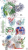
Sequestration of the U2/BS helix by SF3B1 and location of SF3B1 hotspot mutations and the Pladienolide B binding site. (A) The superhelical SF3B1 HEAT domain sequesters the U2/BS helix and acts as a scaffold that binds numerous proteins. Intron nucleotides downstream from the branch site (BS) exit the HEAT domain at repeats H4–H6 and are subsequently bound by the retention and splicing (RES) protein SNU17, followed by the helicase PRP2. SF3B3, which interacts with HEAT repeats H5 and H6 and H19 and H20, has been omitted. (B) Schematic of the SF3B1 HEAT repeats and their interaction partners. Proteins contacting both amino and carboxy-terminal HEAT repeats are colored as in A. (C) Overlapping binding site of Pladienolide B (PB) and the branch site adenosine (BS-A). (Left) Residues of SF3B1 HEAT repeats H15–H17 and PHF5A that form the BS-A binding pocket in the human Bact complex, in which the HEAT domain shows a closed conformation. (Right) The H15–H17 hinge region of SF3B1 bound by PB (orange sticks), which locks the SF3B1 HEAT domain in an open conformation. (D) SF3B1 residues frequently mutated in various human cancers (red lettering) cluster near the 3′ end of the intron where it exits the HEAT superhelix (left). (Right) Space filling model with surface potential (red, negatively charged amino acids; blue, positively charged) of the SF3B1 region shown in the left panel. Many hotspot mutations are nonconserved changes that alter the amino acid’s charge. (E) Meandering path of the SKIP protein (red) in hBact and its numerous interaction partners.
Figure 5.
Figure 5.
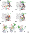
Conformational dynamics of the human Bact complex. (A) Eight major conformational states of the human Bact complex were obtained by computational sorting of images. Only states 2, 4, 6, and 8 are shown. The positions of the most mobile proteins/protein domains are indicated in the electron microscopic (EM) density of Bact. State 4 corresponds to the high-resolution hBact structure from Haselbach et al. (2018) that is shown in Figures 3 and 4. Density for the PRP19HB, PPIL1, and most of PRP17 cannot be recognized in states 1 and 2, even though there is evidence that these proteins, as well as nearly all other hBact proteins, are already present in the Bact complex represented by stage 1. This means that they bind in a highly flexible manner and are simply not discernable by EM, as opposed to being first recruited to hBact at a subsequent step. In state 4, U5-40KWD40 has shifted upward by 2 nm, generating a docking site for one end of the PRP19HB, stabilizing its conformation and leading to its appearance in state 4. Similarly, density for PRP17 and PPIL1 is also first detected in state 4, in which PPIL1 now interacts with PRP19HB. In state 6, PPIL2 is no longer detected, and in state 8, PPIE moves toward PRP17. The U2 3′ domain also moves by several nm from states 2 to 8. (B) Comparison of the spatial organization of the U2 3′ domain, SYF1, SYF3, AQR, PPIE, and the PRP17 WD40 domain in states 2 and 8 of hBact and in the hC complex.
Figure 6.
Figure 6.
Molecular organization of the human C and C* complexes. (A) Overview of the structural organization of the human C and C* complexes. In higher eukaryotes, the exon junction complex (EJC) is deposited 20–24 nucleotides upstream of the exon–exon junction of spliced mRNAs (Le Hir et al. 2000). In hC*, eIF4AIII binds 6 nts of the 5′ exon at a position close to where the EJC should be deposited, and at the same time interacts with MAGOH and MLN51, and U5-SNU114. (B) Interaction of PRP8RH with the U2/BS helix in the C (left) and C* complex (right), as well as interactions of CCDC49 (CWC25), CCDC94 (YJU2) and ISY1 within the RNP core of the C complex (left). (C) Conformational dynamics of the PRP8 RH domain and movements of the U2/BS helix. The position of the branched intron structure formed after step 1 of splicing is stabilized by contacts with PRP8RH and Prp17. The branch site adenosine (BS-A) is indicated by a black circle.
Figure 7.
Figure 7.
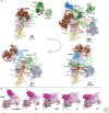
Structural dynamics of the spliceosome. (A) Cartoon depicting the major structural rearrangements that occur during the transitions from the human B to Bact, Bact to C, and C to C* complex. (B) Rearrangements of the Prp8 reverse transcriptase-like (RT) domain, Linker (α-finger), endonuclease-like (En) domain, helical bundle (HB), and amino-terminal (NTD) domain during B, Bact, and C complex formation (indicated by arrows in the preceding complex). Green arrow indicates rotation of HB relative to the RT domain. Black arrow indicates the cleft between the Prp8 NTD and En domains in the Bact complex that are occupied initially by regions of RNF113A and CWC27.

References

    1. Absmeier E, Wollenhaupt J, Mozaffari-Jovin S, Becke C, Lee CT, Preussner M, Heyd F, Urlaub H, Lührmann R, Santos KF, et al. 2015. The large N-terminal region of the Brr2 RNA helicase guides productive spliceosome activation. Genes Dev 29: 2576–2587. - PMC - PubMed
    1. Agafonov DE, Deckert J, Wolf E, Odenwälder P, Bessonov S, Will CL, Urlaub H, Lührmann R. 2011. Semiquantitative proteomic analysis of the human spliceosome via a novel two-dimensional gel electrophoresis method. Mol Cell Biol 31: 2667–2682. - PMC - PubMed
    1. Agafonov DE, Kastner B, Dybkov O, Hofele RV, Liu WT, Urlaub H, Lührmann R, Stark H. 2016. Molecular architecture of the human U4/U6.U5 tri-snRNP. Science 351: 1416–1420. - PubMed
    1. Alsafadi S, Houy A, Battistella A, Popova T, Wassef M, Henry E, Tirode F, Constantinou A, Piperno-Neumann S, Roman-Roman S, et al. 2016. Cancer-associated SF3B1 mutations affect alternative splicing by promoting alternative branchpoint usage. Nat Commun 7: 10615. - PMC - PubMed
    1. Bai R, Yan C, Wan R, Lei J, Shi Y. 2017. Structure of the post-catalytic spliceosome from Saccharomyces cerevisiae. Cell 171: 1589–1598. - PubMed

Publication types