. 2019 Mar 26;116(13):6379-6384.
doi: 10.1073/pnas.1817391116. Epub 2019 Feb 14.

Insulin signaling in the hippocampus and amygdala regulates metabolism and neurobehavior

Affiliations

Insulin signaling in the hippocampus and amygdala regulates metabolism and neurobehavior

Marion Soto et al. Proc Natl Acad Sci U S A. .

Abstract

Previous studies have shown that insulin and IGF-1 signaling in the brain, especially the hypothalamus, is important for regulation of systemic metabolism. Here, we develop mice in which we have specifically inactivated both insulin receptors (IRs) and IGF-1 receptors (IGF1Rs) in the hippocampus (Hippo-DKO) or central amygdala (CeA-DKO) by stereotaxic delivery of AAV-Cre into IRlox/lox/IGF1Rlox/lox mice. Consequently, both Hippo-DKO and CeA-DKO mice have decreased levels of the GluA1 subunit of glutamate AMPA receptor and display increased anxiety-like behavior, impaired cognition, and metabolic abnormalities, including glucose intolerance. Hippo-DKO mice also display abnormal spatial learning and memory whereas CeA-DKO mice have impaired cold-induced thermogenesis. Thus, insulin/IGF-1 signaling has common roles in the hippocampus and central amygdala, affecting synaptic function, systemic glucose homeostasis, behavior, and cognition. In addition, in the hippocampus, insulin/IGF-1 signaling is important for spatial learning and memory whereas insulin/IGF-1 signaling in the central amygdala controls thermogenesis via regulation of neural circuits innervating interscapular brown adipose tissue.

Keywords: amygdala; cognition; hippocampus; insulin; metabolism.


Conflict of interest statement

Conflict of interest statement: C.R.K. and S.G. are coauthors on a 2016 review article.

Figures

Fig. 1.
Fig. 1.
AAV-Cre mediates efficient gene recombination in the hippocampus (Hippo-DKO) and central amygdala (CeA-DKO) of IR/IGF1Rlox/lox mice. (A) Bilateral AAV injection sites of the anterior and posterior hippocampus. Representative images of immunohistochemical staining of GFP and DAPI in the anterior and posterior hippocampus. (B) Immunoblotting of IR and IGF1R in the hippocampus of adult IR/IGF1Rlox/lox mice injected with AAV-GFP or AAV-Cre-GFP. Bottom, densitometry analysis. (C) Bilateral AAV injection sites of the central amygdala. Representative images of immunohistochemical staining of GFP and DAPI in the central amygdala. (D) Immunoblotting of IR and IGF1R in the central amygdala of adult IR/IGF1Rlox/lox mice injected with AAV-GFP or AAV-Cre-GFP. Bottom, densitometry analysis. *P < 0.05 by unpaired t test, n = 6. Data are presented as mean ± SEM.
Fig. 2.
Fig. 2.
Decreased expression of glutamate receptor 1 in the synaptosome of Hippo-DKO and CeA-DKO mice. (A and B) Representative Western blots and quantification of glutamate receptor 1 and 2 (GluA1 and GluA2) in synaptosomes extracted from the hippocampus in Hippo-DKO and control mice (A) and the central amygdala in CeA-DKO and control mice (B). GAPDH serve as loading controls. *P < 0.05, **P < 0.01 by unpaired t test, n = 6. Quantitative data are presented as mean ± SEM.
Fig. 3.
Fig. 3.
Glucose homeostasis is regulated by IR/IGF1R signaling in the hippocampus and central amygdala. (A) Blood glucose levels in the random-fed state of Hippo-DKO (n = 31) or Hippo-CTR mice (n = 27). (B) Oral glucose tolerance test (OGTT) performed in Hippo-DKO or Hippo-CTR mice (n =13). (C) Blood glucose levels in the random-fed state of CeA-DKO (n = 29) or CeA-CTR mice (n = 28). (D) OGTT performed in CeA-DKO or CeA-CTR mice (n = 6). *P < 0.05, **P < 0.01, ***P < 0.001 by unpaired t test. Data are presented as mean ± SEM. AUC, area under the curve.
Fig. 4.
Fig. 4.
IR/IGF1R signaling in the amygdala contributes to cold-induced thermogenesis. (A) Rectal temperature in Hippo-CTR (n = 4) and Hippo-DKO mice (n = 5) during a 3-h exposure to a 6 °C environment. (B) Rectal temperature in CeA-CTR (n = 9) and CeA-DKO mice (n = 10) during a 3-h exposure to a 6 °C environment. *P < 0.05 by unpaired t test. (C) Thermal images using a FLIR T300 Infrared Camera showing surface temperature after 3 h at 6 °C between CeA-CTR and CeA-DKO mice. (D) Fluorescent images of brain sections of mice 7 d after injection of the PRV-765 virus in the BAT. BLA, basolateral amygdalar area; BMA, basomedial amygdalar nucleus; COA, cortical amygdalar area; ENTl, entorhinal area lateral part; PA, piriform-amygdalar area; PIA, piriform area; TR, postpiriform transition area. Data are presented as mean ± SEM.
Fig. 5.
Fig. 5.
Hippo-DKO and CeA-DKO mice display increased anxiety-like behaviors. (A) Time spent in the center zone and entries in center zone in open field test in Hippo-CTR (n = 12) and Hippo-DKO mice (n = 15). (B) Assessment of anxiety as number of buried marbles over 30 min during a marble-burying task in Hippo-CTR (n = 15) and Hippo-DKO mice (n = 17). (C) Time spent in light compartment during light/dark box test in Hippo-CTR (n = 11) and Hippo-DKO mice (n = 12). (D) Time spent in the center zone and entries in center zone in open field test in CeA-CTR (n = 13) and CeA-DKO mice (n = 14). (E) Assessment of anxiety as number of buried marbles over 30 min during a marble-burying task test in CeA-CTR (n = 9) and CeA-DKO mice (n = 9). (F) Time spent in light compartment during light/dark box test in CeA-CTR (n = 18) and CeA-DKO mice (n = 19). *P < 0.05, **P < 0.01 by unpaired t test. Data are presented as mean ± SEM.
Fig. 6.
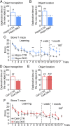
Fig. 6.
Hippo-DKO and CeA-DKO mice have impaired cognition. (A) Time spent exploring the novel object during the test session of the object recognition task in Hippo-CTR (n = 8) and Hippo-DKO mice (n = 9). (B) Time spent exploring the object that was moved during the test session of the object location task in Hippo-CTR (n = 4) and Hippo-DKO mice (n = 5). (C) Errors recorded during the 10 acquisition trials, 1-wk, and 1-mo memory testing of the Stone T maze in Hippo-CTR (n = 7) and Hipp-DKO mice (n = 8). (D) Time spent exploring the novel object during the test session of the object recognition task in CeA-CTR (n = 9) and CeA-DKO mice (n = 10). (E) Time spent exploring the object that was moved during the test session of the object location task in CeA-CTR (n = 6) and CeA-DKO mice (n = 6). (F) Errors recorded during the 10 acquisition trials, 1-wk, and 1-mo memory testing of the Stone T maze in CeA-CTR (n = 7) and CeA-DKO mice (n = 9). #P < 0.05, ##P < 0.01, ###P < 0.001 by unpaired t test. *P < 0.05, **P < 0.01 between DKO and control mice by unpaired t test. Data are presented as mean ± SEM.

Comment in

References

    1. Kahn CR, Freychet P, Roth J, Neville DM., Jr Quantitative aspects of the insulin-receptor interaction in liver plasma membranes. J Biol Chem. 1974;249:2249–2257. - PubMed
    1. Accili D, et al. Early neonatal death in mice homozygous for a null allele of the insulin receptor gene. Nat Genet. 1996;12:106–109. - PubMed
    1. Steele-Perkins G, et al. Expression and characterization of a functional human insulin-like growth factor I receptor. J Biol Chem. 1988;263:11486–11492. - PubMed
    1. Liu JP, Baker J, Perkins AS, Robertson EJ, Efstratiadis A. Mice carrying null mutations of the genes encoding insulin-like growth factor I (Igf-1) and type 1 IGF receptor (Igf1r) Cell. 1993;75:59–72. - PubMed
    1. DeChiara TM, Efstratiadis A, Robertson EJ. A growth-deficiency phenotype in heterozygous mice carrying an insulin-like growth factor II gene disrupted by targeting. Nature. 1990;345:78–80. - PubMed

Publication types

MeSH terms