. 2019 Feb 14;9(1):2124.
doi: 10.1038/s41598-018-38099-w.

Numerical Simulations of Realistic Lead Trajectories and an Experimental Verification Support the Efficacy of Parallel Radiofrequency Transmission to Reduce Heating of Deep Brain Stimulation Implants during MRI

Affiliations

Numerical Simulations of Realistic Lead Trajectories and an Experimental Verification Support the Efficacy of Parallel Radiofrequency Transmission to Reduce Heating of Deep Brain Stimulation Implants during MRI

C E McElcheran et al. Sci Rep. .

Abstract

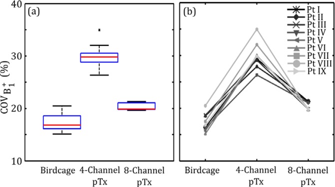
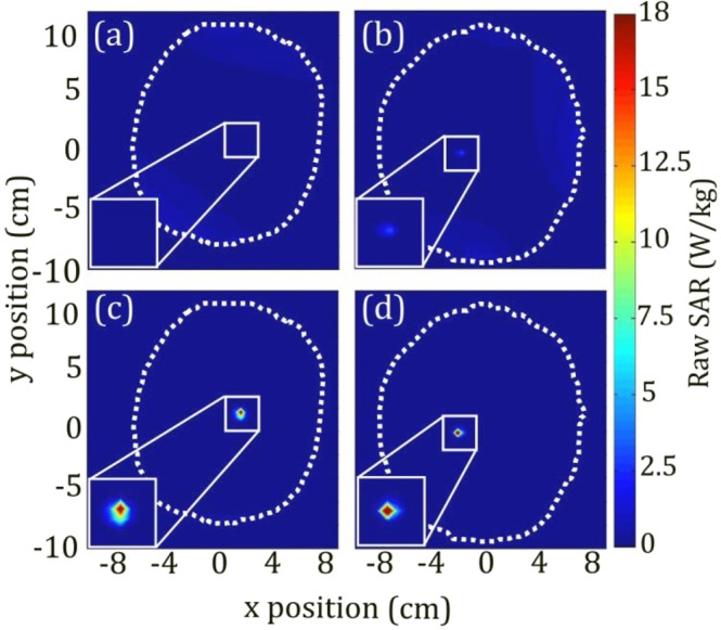
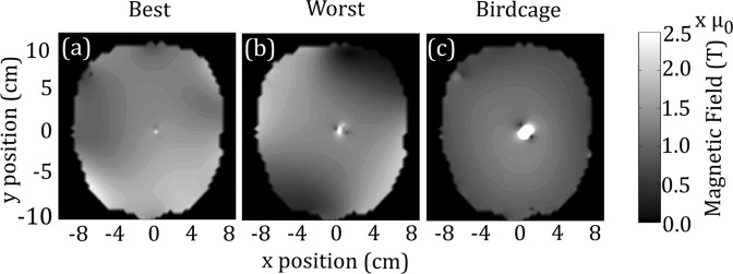
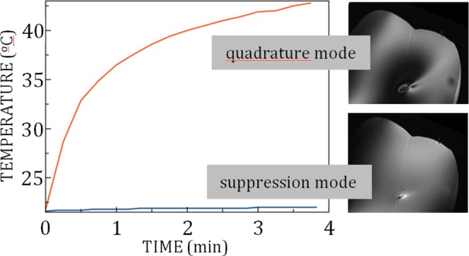
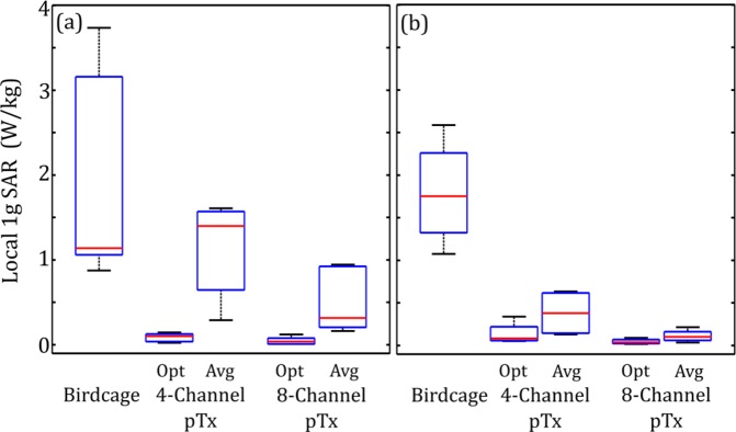
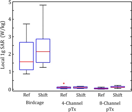
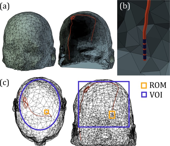
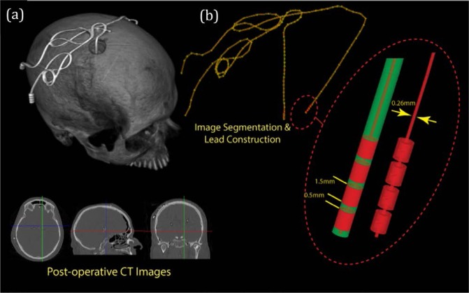
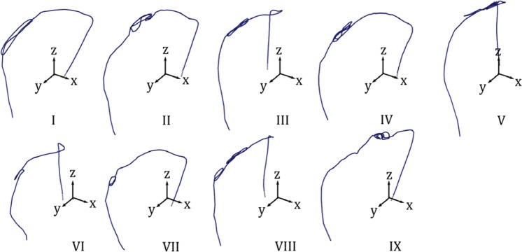
Patients with deep brain stimulation (DBS) implants may be subject to heating during MRI due to interaction with excitatory radiofrequency (RF) fields. Parallel RF transmit (pTx) has been proposed to minimize such RF-induced heating in preliminary proof-of-concept studies. The present work evaluates the efficacy of pTx technique on realistic lead trajectories obtained from nine DBS patients. Electromagnetic simulations were performed using 4- and 8-element pTx coils compared with a standard birdcage coil excitation using patient models and lead trajectories obtained by segmentation of computed tomography data. Numerical optimization was performed to minimize local specific absorption rate (SAR) surrounding the implant tip while maintaining spatial homogeneity of the transmitted RF magnetic field (B1+), by varying the input amplitude and phase for each coil element. Local SAR was significantly reduced at the lead tip with both 4-element and 8-element pTx (median decrease of 94% and 97%, respectively), whereas the median coefficient of spatial variation of B1+ inhomogeneity was moderately increased (30% for 4-element pTx and 20% for 8-element pTx) compared to that of the birdcage coil (17%). Furthermore, the efficacy of optimized 4-element pTx was verified experimentally by imaging a head phantom that included a wire implanted to approximate the worst-case lead trajectory for localized heating, based on the simulations. Negligible temperature elevation was observed at the lead tip, with reasonable image uniformity in the surrounding region. From this experiment and the simulations based on nine DBS patient models, optimized pTx provides a robust approach to minimizing local SAR with respect to lead trajectory.


Conflict of interest statement

The authors declare no competing interests.

Figures

Figure 1
Figure 1
Estimated local 1 g SAR in the ROM surrounding the tip of the DBS electrode for lead trajectories obtained from nine different patients. (a) Box and whisker plot showing median, interquartile range (IQR) and data range. Data lying outside 1.5•IQR are shown as outliers. (b) Line plot of the individual patient-trajectory data. (c) Corresponding peak temperature increase at the tip of the DBS electrode. Results are shown for the birdcage coil operated in quadrature transmission mode and for optimized 4-channel and 8-channel pTx.
Figure 2
Figure 2
B1+ inhomogeneity in the VOI for lead trajectories obtained from nine different patients. Results are shown for the birdcage coil operated in quadrature transmission mode and for optimized 4-channel and 8-channel pTx. (a) Box and whisker plot showing median, IQR and data range. Data lying outside 1.5•IQR are shown as outliers. (b) Line plot of the individual patient-trajectory data.
Figure 3
Figure 3
Estimated whole-head averaged SAR for lead trajectories obtained from nine different patients. Results are shown for the birdcage coil operated in quadrature transmission mode and for optimized 4-channel and 8-channel pTx. (a) Box and whisker plot showing median, interquartile range and data range. (b) Line plot of the individual patient-trajectory data.
Figure 4
Figure 4
Contour plot of raw SAR (resolution: 0.39 cm × 0.45 cm × 0.35 cm) for (a) best solution for local 1 g SAR and (b) worst solution for local 1 g SAR with corresponding SAR plots for birdcage excitation (c,d). White dotted line indicates outline of patient head. Position of plane in z-direction corresponds with maximum SAR location and may differ between plots. Implant region shown at 2x magnification in bottom left corner.
Figure 5
Figure 5
Contour plots of (a) best solution for B1+ inhomogeneity, (b) worst solution for B1+ inhomogeneity, and (c) quadrature birdcage excitation.
Figure 6
Figure 6
Plots of temperature versus time for pTx TSE MRI in quadrature mode and in suppression mode, recorded at the exposed tip of a wire implanted in a uniform head phantom with a trajectory approximating a high-risk DBS scenario. Images are also shown at the slice location intersecting the wire tip, to evaluate signal uniformity and RF coupling artifacts. Shading at the left side of each image is due to extraneous application of an RF saturation band pulse, not RF coupling. The thin diagonal line shown in both images arose from filling the phantom in two pouring sessions.
Figure 7
Figure 7
Estimated local 1 g SAR in the ROM surrounding the tip of the DBS electrode for lead trajectories obtained from nine different patients. Results are shown for the birdcage coil operated in quadrature transmission mode and for 4-channel and 8-channel pTx excited with optimized, patient-specific inputs (Opt) and amplitude and phase shifts averaged over patients with the same lateralization (Avg). The box and whisker plots show median, interquartile range and data range for patients with (a) left-lateralized implants and (b) right-lateralized implants.
Figure 8
Figure 8
Box and whisker plots showing median, interquartile range (IQR) and data range of estimated local 1 g SAR in the ROM surrounding the tip of the DBS electrode for lead trajectories obtained from nine different patients. Results are shown for original patient-trajectory model orientations and for patient-trajectory models after random rigid-body shift, for the birdcage coil operated in quadrature transmission mode and for optimized 4-channel and 8-channel pTx. Data lying outside 1.5•IQR are shown as outliers.
Figure 9
Figure 9
(a) Example patient head mesh model with implanted DBS lead trajectory. (b) Zoomed-in view of the DBS target location showing wire insulation in red and four exposed electrodes in dark blue. (c) Transparent view of the model showing the region of minimization (ROM) for minimizing local power deposition (yellow) and the volume of interest (VOI) for minimizing B1+ inhomogeneity (blue.).
Figure 10
Figure 10
Coil configurations for (a) birdcage excitation, (b) 4-element pTx excitation and (c) 8-element pTx excitation. The Cartesian coordinate system (red) and an example of a patient model are also shown.
Figure 11
Figure 11
(a) Three-dimensional reconstruction of intra-operative CT images from a DBS patient, with representative axial, coronal and sagittal images shown below. (b) Segmentation of DBS lead, with zoomed-in region showing in detail how the lead tip was modelled.
Figure 12
Figure 12
Lead trajectories for nine patient models (patient numbers I–IX).

References

    1. Chen XL, Xiong YY, Xu GL, Liu XF. Deep brain stimulation. Interventional neurology. 2013;1:200–212. doi: 10.1159/000353121. - DOI - PMC - PubMed
    1. Blomstedt P, Hariz G-M, Hariz MI, Koskinen L-O. Thalamic deep brain stimulation in the treatment of essential tremor: a long-term follow-up. British journal of neurosurgery. 2007;21:504–509. doi: 10.1080/02688690701552278. - DOI - PubMed
    1. Flora ED, Perera CL, Cameron AL, Maddern GJ. Deep brain stimulation for essential tremor: a systematic review. Movement disorders. 2010;25:1550–1559. doi: 10.1002/mds.23195. - DOI - PubMed
    1. Ostrem JL, Starr PA. Treatment of dystonia with deep brain stimulation. Neurotherapeutics. 2008;5:320–330. doi: 10.1016/j.nurt.2008.01.002. - DOI - PMC - PubMed
    1. Vercueil L, et al. Deep brain stimulation in the treatment of severe dystonia. Journal of neurology. 2001;248:695–700. doi: 10.1007/s004150170116. - DOI - PubMed