. 2019 Feb 14;9(1):2033.
doi: 10.1038/s41598-018-36578-8.

Antibody-mediated biorecognition of myelin oligodendrocyte glycoprotein: computational evidence of demyelination-related epitopes

Affiliations

Antibody-mediated biorecognition of myelin oligodendrocyte glycoprotein: computational evidence of demyelination-related epitopes

Jéssica Cristiane Magalhães Ierich et al. Sci Rep. .

Abstract

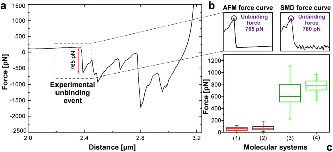
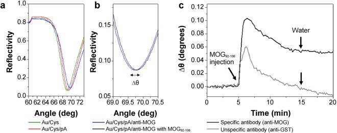
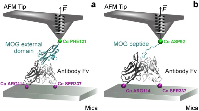
Antigen-antibody interaction is crucial in autoimmune disease pathogenesis, as multiple sclerosis and neuromyelitis optica. Given that, autoantibodies are essential biomolecules, of which the myelin oligodendrocyte glycoprotein (MOG) can figure as a target. Here we combined Molecular Dynamics (MD), Steered Molecular Dynamics (SMD), and Atomic Force Microscope (AFM) to detail MOG recognition by its specific antibody. The complex model consisted of the MOG external domain interacting with an experimental anti-MOG antibody from the Protein Data Bank (1PKQ). Computational data demonstrated thirteen MOG residues with a robust contribution to the antigen-antibody interaction. Comprising five of the thirteen anchor residues (ASP102, HIS103, SER104, TYR105, and GLN106), the well-known MOG92-106 peptide in complex with the anti-MOG was analysed by AFM and SMD. These analyses evidenced similar force values of 780 pN and 765 pN for computational and experimental MOG92-106 and anti-MOG detachment, respectively. MOG92-106 was responsible for 75% of the total force measured between MOG external domain and anti-MOG, holding the interaction with the antibody. The antigen-antibody binding was confirmed by Surface Plasmon Resonance (SPR) measurements. Combined approaches presented here can conveniently be adjusted to detail novel molecules in diseases research. This can optimize pre-clinical steps, guiding experiments, reducing costs, and animal model usage.


Conflict of interest statement

The authors declare no competing interests.

Figures

Figure 1
Figure 1
Evolution of system biomolecules RMSD values during the simulation time from the initial structure. (a) Complex, Fab, and MOG RMSD values calculated during the entire simulation. (b) RMSD evolution of each Fab region as follows: variable region, CDR, and constant region.
Figure 2
Figure 2
Hydrogen bond contribution for antigen-antibody complex formation. (a) The complex formed by the MOG external domain (green) and the anti-MOG Fab portion (light chain highlighted in white and heavy chain in grey). (b) Identification of the six CDR in the anti-MOG Fab structure, in which L1 is shown in red, L2 in blue, L3 in orange, H1 in yellow, H2 in cyan, and H3 in purple. (c) Contributions of CDR residues in terms of hydrogen bonds (black dashed lines) in the interaction between MOG (green) and anti-MOG Fab for every 40 ns of simulation. (d) Hydrogen bonds quantified for the complex in every 10 ns of simulation.
Figure 3
Figure 3
Electrostatic potential (−3.0 KBT/e to + 3.0 KBT/e) of the MOG-Fab complex. (a) The complex structure composed of MOG external domain (green) and the anti-MOG Fab portion (black). (b) Complex electrostatic potential representation in which the interaction site is represented by the horizontal plane. (c) Superior view of the horizontal plane with the most interactive region of the MOG molecule comprised in the dark square. (d) Inferior view of the horizontal plane with the most interactive region of the anti-MOG Fab portion comprised in the dark square.
Figure 4
Figure 4
Computational forces involved in the antigen-antibody complex. (a) SMD force curve of the interaction between MOG external domain and the anti-MOG Fv portion. (b) SMD force curve of the interaction between the MOG92–106 peptide and the anti-MOG Fv portion.
Figure 5
Figure 5
Interaction between MOG immunogenic peptide and antibody regarding force values. (a) AFM force curve of the complex formed by MOG92–106 and rabbit anti-MOG IgG. (b) Obtained AFM force curve in comparison with SMD data. (c) Boxplot of the AFM and SMD data including control measurements.
Figure 6
Figure 6
(a) SPR angular scan of the Au/Cys/pA/anti-MOG sensing surface assembly: Au/Cys in green, Au/Cys/pA in red, and Au/Cys/pA/anti-MOG (sensing surface) in blue; (b) SPR angle shift Δθ due to the specific interaction between the sensing surface in blue with MOG92–106 peptide in black. (c) Real-time Δθ monitoring during the MOG92–106 followed by water injection over the specific anti-MOG in black and unspecific anti-GST surface in grey.
Figure 7
Figure 7
Schematic model of the SMD simulated systems with AFM tip and mica substrate indicated in representative positions. (a) SMD input of the complex MOG-Fv. (b) SMD input of the complex MOG92–106-Fv.

References

    1. Sela-Culang I, Kunik V, Ofran Y. The structural basis of antibody-antigen recognition. Front. Immunol. 2013;4:302. doi: 10.3389/fimmu.2013.00302. - DOI - PMC - PubMed
    1. Marillet, S., Lefranc, M.-P., Boudinot, P. & Cazals, F. Novel structural parameters of ig-ag complexes yield a quantitative description of interaction specificity and binding affinity. Front. Immunol. 8 (2017). - PMC - PubMed
    1. Fukuda N, et al. Role of the mobility of antigen binding site in high affinity antibody elucidated by surface plasmon resonance. J. Biochem. 2017;161:37–43. doi: 10.1093/jb/mvw050. - DOI - PubMed
    1. Kinzel, S. & Weber, M. S. The role of peripheral CNS-directed antibodies in promoting inflammatory CNS demyelination. Brain Sci. 7 (2017). - PMC - PubMed
    1. Mayer MC, Meinl E. Glycoproteins as targets of autoantibodies in CNS inflammation: MOG and more. Ther. Adv. Neurol. Disord. 2012;5:147–159. doi: 10.1177/1756285611433772. - DOI - PMC - PubMed

Publication types

MeSH terms