The evolution of the anterior capsulotomy
- PMID: 30766624
- PMCID: PMC6372876
- DOI: 10.5114/wiitm.2019.81313
The evolution of the anterior capsulotomy
Abstract
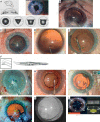
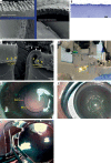
The paper describes the development of the anterior capsulotomy from its early crude beginnings in the 18th century to the possibility of automated surgery today via continuous curvilinear capsulorhexis (CCC). The reasons for the opening of the capsule have changed from a roughly made tear to allow access to the nucleus for its extraction, to the creation of more regular openings to allow support for intraocular lenses. With the development of continuous circular tears it was possible to be certain to contain the intraocular lens (IOL) in the capsular bag. In recent times we have the ability to achieve precision in size and location with lasers and other technologies. This means the capsulotomy can be used to hold the IOL, which will improve the centration of the optic. This is important in premium lenses and should improve predictability of the effective lens position. All of these changes will be highlighted with appropriate illustrations.
Keywords: anterior capsulotomy; cataract; continuous curvilinear capsulorhexis; phacoemulsification.
Conflict of interest statement
Richard Packard is a consultant to Excel-Lens (USA), which produces CAPSULaser. The other authors declare that there is no conflict of interest regarding the publication of this manuscript.
Figures


References
-
- Gimbel H, Neuhann T. Development, advantages, and methods of the continuous circular capsulorhexis technique. J Cataract Refract Surg. 1990;16:31–7. - PubMed
-
- Haeussler-Sinangin Y, Dahlhoff D, Schultz T, Dick HB. Clinical performance in continuous curvilinear capsulorhexis creation supported by a digital image guidance system. J Cataract Refract Surg. 2017;43:348–52. - PubMed
-
- Hollick EJ, Spalton DJ, Meacock WR. The effect of capsulorhexis size on posterior capsular opacification: one-year results of a randomized prospective trial. Am J Ophthalmol. 1999;128:271–9. - PubMed
-
- Popovic M, Campos-Muller X, Schlenker M, Ahmed I. Efficacy and safety of femtosecond laser-assisted cataract surgery compared with manual cataract surgery: a meta-analysis of 14 567 eyes. Ophthalmology. 2016;123:2113–26. - PubMed
-
- Mursch-Edlmayr A, Bolz M, Luft N, et al. Intraindividual comparison between femtosecond laser-assisted and conventional cataract surgery. J Cataract Refract Surg. 2017;43:215–22. - PubMed
Publication types
LinkOut - more resources
Full Text Sources
