

doi: 10.1016/j.vgie.2018.09.016.
eCollection 2019 Feb.
Recalcitrant embedded biliary self-expanding metal stents: a novel technique for endoscopic extraction
Affiliations
- PMID: 30766947
- PMCID: PMC6362311
- DOI: 10.1016/j.vgie.2018.09.016
Item in Clipboard
Recalcitrant embedded biliary self-expanding metal stents: a novel technique for endoscopic extraction
VideoGIE.
.
No abstract available
Keywords: ERC, endoscopic retrograde cholangioscopy; SEMSs, self-expanding metal stents; cSEMSs, covered SEMSs; uSEMSs, uncovered SEMSs.
Figures

Coronal CT image from the portal venous phase, performed at admission, showing a diffusely abnormal-appearing gallbladder with wall thickening and a soft-tissue mass in the central hilar region associated with intrahepatic bile duct dilatation.

MRCP at initial presentation demonstrating a 2-cm stricture in the common hepatic duct with bilateral intrahepatic biliary dilatation.

Coronal image from 5-minute delayed-phase magnetic resonance imaging 5 weeks after presentation showing nearly complete resolution of the previously observed findings.

The distal end of the uncovered SEMS is clearly visible in the duodenum. SEMS, Self-expanding metal stent.

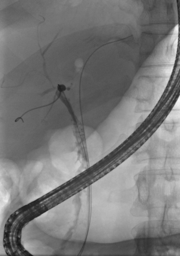
Passage of a guidewire through 2 fenestrations in the uncovered stent, catching the distal end in a loop.

An uncovered SEMS with the positioned guidewire and a Soehendra lithotriptor in place. SEMS, Self-expanding metal stent.

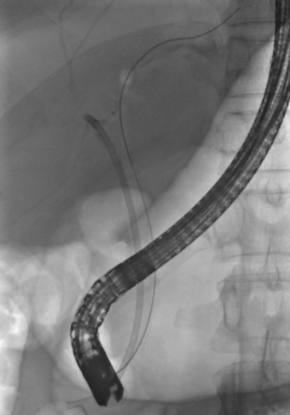
Distortion of distal stent due to snapping of the stent wire through which the guidewire was looped as the wire was pulled into the sheath.
Postprocedure cholangiogram demonstrating an intact and patent biliary tree with air in the extrahepatic bile ducts and left intrahepatic duct.
The right-sided stent is in place with the guidewire within the left duct for subsequent placement of the left-sided stent.
References
-
- Dumonceau J.M., Tringali A., Blero D. Biliary stenting: indications, choice of stents and results: European Society of Gastrointestinal Endoscopy (ESGE) clinical guideline. Endoscopy. 2012;44:277–298. - PubMed
-
- Kullman E., Frozanpor F., Soderlund C. Covered versus uncovered self-expandable nitinol stents in the palliative treatment of malignant distal biliary obstruction: results from a randomized, multicenter study. Gastrointest Endosc. 2010;72:915–923. - PubMed
-
- Lee S.J., Kim M.D., Lee M.S. Comparison of the efficacy of covered versus uncovered metallic stents in treating inoperable malignant common bile duct obstruction: a randomized trial. J Vasc Interv Radiol. 2014;25:1912–1920. - PubMed
-
- Chahal P., Baron T.H. Expandable metal stents for endoscopic bilateral stent-within-stent placement for malignant hilar biliary obstruction. Gastrointest Endosc. 2010;71:195–199. - PubMed
-
- Maccioni F., Rossi M., Salvatori F.M. Metallic stents in benign biliary strictures: three-year follow-up. Cardiovasc Intervent Radiol. 1992;15:360–366. - PubMed
LinkOut - more resources
Full Text Sources
