. 2019 Jan 2;3(2):262-276.
doi: 10.1002/hep4.1302. eCollection 2019 Feb.

Impaired Hepatic Phosphatidylcholine Synthesis Leads to Cholestasis in Mice Challenged With a High-Fat Diet

Affiliations

Impaired Hepatic Phosphatidylcholine Synthesis Leads to Cholestasis in Mice Challenged With a High-Fat Diet

Sereana Wan et al. Hepatol Commun. .

Abstract

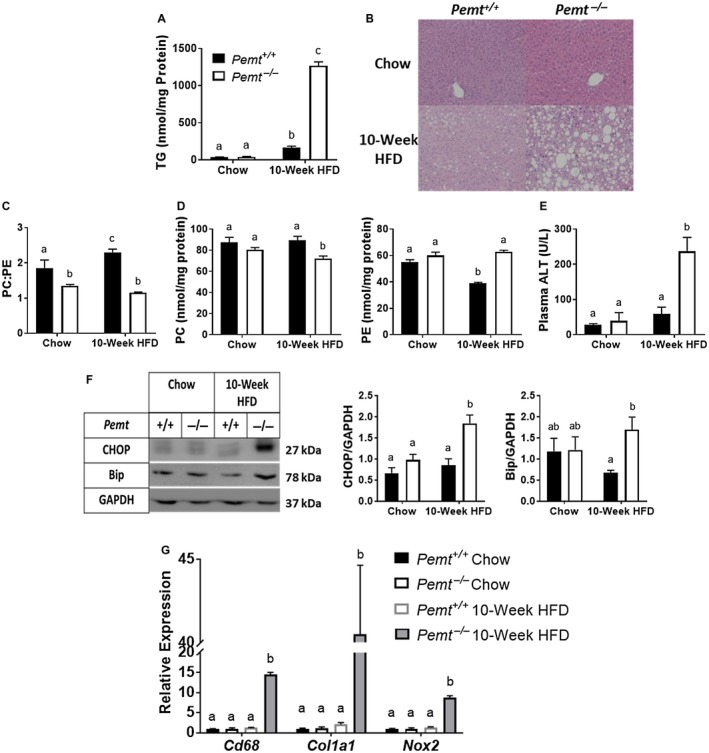
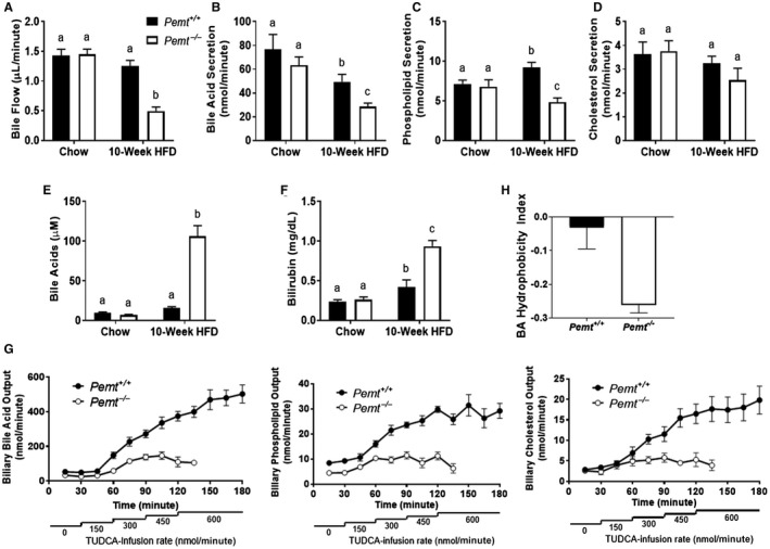
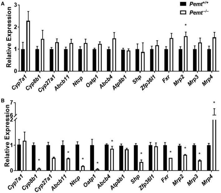
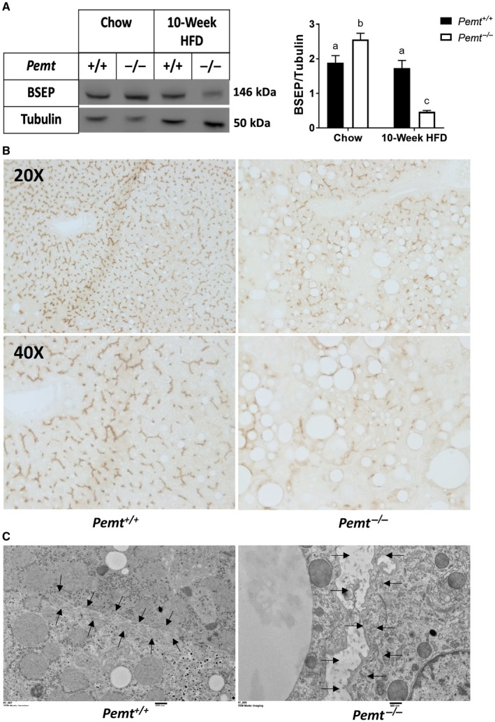
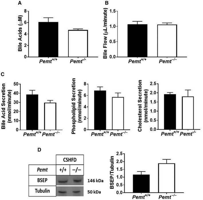
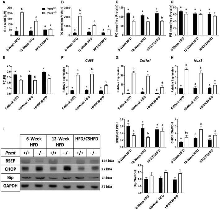
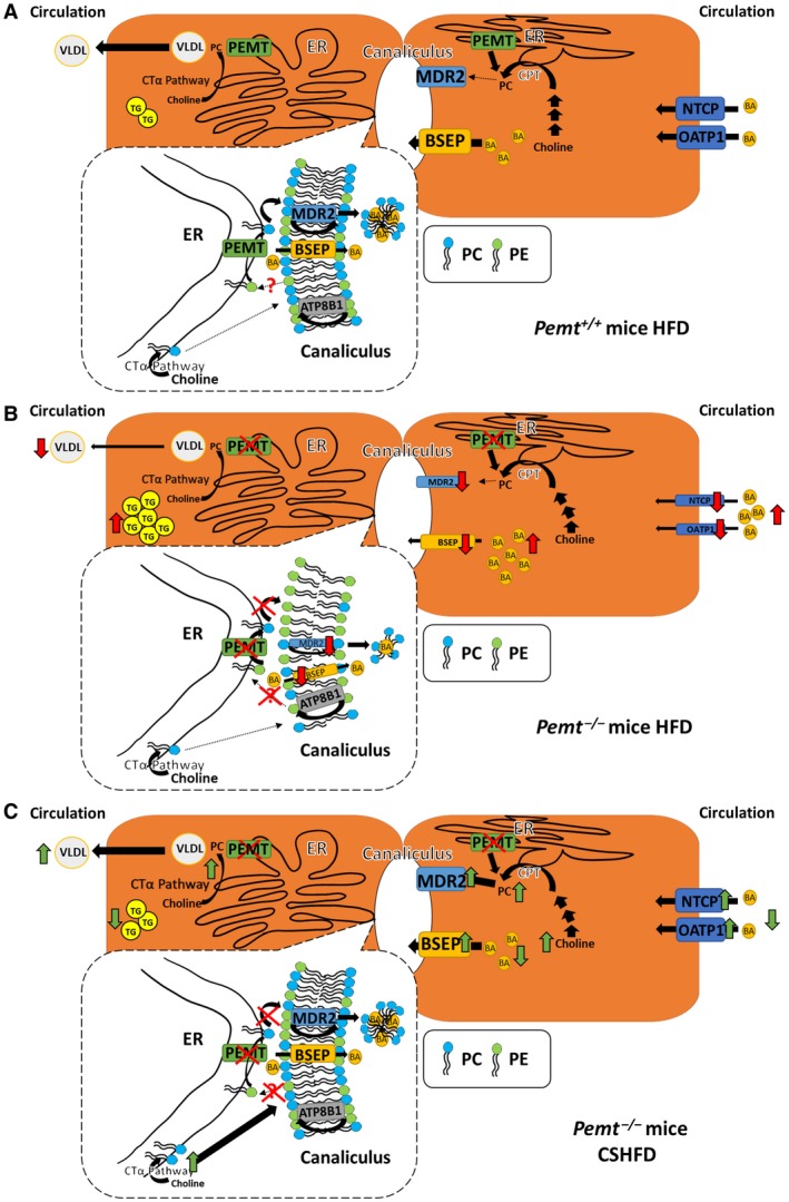
Phosphatidylethanolamine N-methyltransferase (PEMT) is a hepatic integral membrane protein localized to the endoplasmic reticulum (ER). PEMT catalyzes approximately 30% of hepatic phosphatidylcholine (PC) biosynthesis. Pemt-/- mice fed a high-fat diet (HFD) develop steatohepatitis. Interestingly, portions of the ER located close to the canaliculus are enriched in PEMT. Phospholipid balance and asymmetrical distribution by adenosine triphosphatase phospholipid transporting 8B1 (ATP8B1) on the canalicular membrane is required for membrane integrity and biliary processes. We hypothesized that PEMT is an important supplier of PC to the canaliculus and that PEMT activity is critical for the maintenance of canalicular membrane integrity and bile formation following HFD feeding when there is an increase in overall hepatic PC demand. Pemt+/+ and Pemt-/- mice were fed a chow diet, an HFD, or a choline-supplemented HFD. Plasma and hepatic indices of liver function and parameters of bile formation were determined. Pemt-/- mice developed cholestasis, i.e, elevated plasma bile acid (BA) concentrations and decreased biliary secretion rates of BAs and PC, during HFD feeding. The maximal BA secretory rate was reduced more than 70% in HFD-fed Pemt-/- mice. Hepatic ABCB11/bile salt export protein, responsible for BA secretion, was decreased in Pemt-/- mice and appeared to be retained intracellularly. Canalicular membranes of HFD-fed Pemt-/- mice contained fewer invaginations and displayed a smaller surface area than Pemt+/+ mice. Choline supplementation (CS) prevented and reversed the development of HFD-induced cholestasis. Conclusion: We propose that hepatic PC availability is critical for bile formation. Dietary CS might be a potential noninvasive therapy for a specific subset of patients with cholestasis.


Figures

Figure 1
Figure 1
Pemt–/– mice develop NASH when fed the HFD. Pemt+/+ and Pemt–/– mice were fed the chow diet or the HFD for 10 weeks. (A) Hepatic TG mass. (B) Representative hematoxylin and eosin staining of livers of Pemt+/+and Pemt–/– mice fed either the chow diet or the HFD for 10 weeks (magnification ×20). (C) PC:PE molar ratio. (D) Mass of PC and PE. (E) Plasma ALT levels. Values are means ± SEM (n = 6 per group). (F) Bip and ER stress‐responsive CHOP and densitometry. Values are means ± SEM (n = 4 per group). (G) mRNA expression of genes involved in inflammation (Cd68), fibrosis (Col1a1), and oxidative stress (Nox2). Values are means ± SEM (n = 5 per group) and are expressed relative to Pemt+/+ mice fed the chow diet; 2‐way ANOVA, followed by Fisher’s LSD post hoc test. Values that do not share a letter are significantly different (P < 0.05).
Figure 2
Figure 2
Pemt–/– mice develop diet‐induced cholestasis. Pemt+/+ and Pemt–/– mice were fed the chow diet or the HFD for 10 weeks. (A) Basal biliary bile flow and (B) secretion of BAs, (C) phospholipids, and (D) cholesterol. (E) Plasma BAs and (F) total bilirubin concentration. (G) Maximal biliary secretion of BAs, phospholipids, and cholesterol after TUDCA infusion in mice fed the HFD for 10 weeks. (H) Hydrophobicity of the BA pool after 10 weeks of the HFD. Values are means ± SEM (n = 6 per group); 2‐way ANOVA, followed by Fisher’s LSD post hoc test. Values that do not share a letter are significantly different (P < 0.05).
Figure 3
Figure 3
Dysregulation of expression of hepatic genes involved in BA homeostasis in Pemt–/– mice. Hepatic mRNA levels of genes involved in BA synthesis, biliary secretion and basolateral import of BAs, biliary secretion of phospholipids, regulators of BA synthesis, BA homeostasis, and the alternative pathway of BA secretion into bile and circulation were determined in Pemt+/+ and Pemt–/– mice fed (A) chow or (B) the HFD and normalized to cyclophilin mRNA levels. Values are expressed relative to Pemt+/+ mice on the same diet. Values are means ± SEM (n = 5 per group). Student t test; *P < 0.05.
Figure 4
Figure 4
Loss of canalicular structure and BSEP deficiency in Pemt–/– mice. (A) Representative immunoblot of hepatic BSEP protein and quantification in Pemt+/+ and Pemt–/– mice fed the chow diet or the HFD for 10 weeks. Values are means ± SEM (n = 6 per group); 2‐way ANOVA followed by Fisher’s LSD post hoc test. Values that do not share a letter are significantly different (P < 0.05). (B) Representative immunohistochemistry (above, magnification ×20; below, magnification ×40) for BSEP. (C) Electron microscopy of the canalicular membrane (magnification ×6,000) in livers of Pemt+/+ and Pemt–/– mice fed the HFD for 10 weeks. Arrows outline the edge of the canaliculus.
Figure 5
Figure 5
CS prevents cholestasis in Pemt–/– mice. Pemt+/+ and Pemt–/– mice were fed the CSHFD for 10 weeks. (A) Plasma BA concentration. (B) Basal biliary bile flow and (C) BA, phospholipid, and cholesterol secretion. (D) Hepatic BSEP protein and quantification relative to the amount of tubulin. Values are means ± SEM (n = 6 per group); 2‐way ANOVA followed by Fisher’s LSD post hoc test. Values that do not share a letter are significantly different (P < 0.05).
Figure 6
Figure 6
CS improves liver health and treats cholestasis in Pemt–/– mice. Pemt+/+ and Pemt–/– mice were fed the HFD for 6 weeks and either continued on the HFD or the CSHFD for an additional 6 weeks. (A) Plasma BA concentration. (B) Hepatic TG, (C) PC, (D) PE, and (E) PC:PE ratio. Values are means ± SEM (n = 6 per group). mRNA expression of hepatic genes involved in (F) inflammation, (G) fibrosis, and (H) oxidative stress. Values are means ± SEM (n = 5 per group). (I) Hepatic BSEP, CHOP, and Bip and quantification relative to the amount of GAPDH. Values are means ± SEM (n = 6 per group); two‐way ANOVA followed by Fisher’s LSD post‐hoc test. Values that do not share a letter are significantly different (P < 0.05).
Figure 7
Figure 7
Working model of cholestasis in Pemt+/+ and Pemt–/– mice fed the HFD. (A) When Pemt+/+ mice are fed the HFD, BAs are secreted from the hepatocyte into bile by BSEP and removed from the portal circulation by NTCP and OATP1. Hepatic PC is synthesized either by means of the choline pathway or the PEMT pathway (conversion from PE to PC) in the ER. Hepatic PC and TG are secreted into the circulation as VLDL, and PC is secreted into bile. MDR2 flips PC from the inner to the outer membrane of the canalicular membrane from where the PC is extracted into bile by the BAs. ATP8B1 flips PE from the outer to the inner canalicular membrane. The dotted box represents enlargement of the area of ER close to the canaliculus. ER domains that are close to the canalicular membrane are enriched with PEMT, thereby providing PC directly to the canalicular membrane for biliary secretion. In addition, PEMT might deplete PE from the canaliculus so that an appropriate PC:PE ratio is maintained. Moreover, PC is made in areas of the ER not close to the canaliculus through the choline pathway and might also provide PC for biliary secretion. (B) When Pemt–/– mice are fed the HFD, the amount of mRNAs encoding NTCP, OATP1, and BSEP is decreased (mechanism not known), leading to the accumulation of BAs in plasma and, likely, in the liver/hepatocytes. Moreover, the amount of MDR2 is decreased in an attempt to conserve hepatic PC. Thus, because PC is synthesized only by the choline pathway in Pemt–/– hepatocytes, a reduction in hepatic PC leads to impaired VLDL secretion and consequently hepatic steatosis. The lack of PEMT in the hepatocytes reduces the local supply of PC for the canalicular membrane, which cannot be compensated by PC synthesized in the bulk of the ER by the choline pathway. The deficiency of PC in the canalicular membrane would compromise integrity of the canalicular membrane and change in structure of the canaliculus. Arrows in red indicate differences compared to Pemt+/+ mice. (C) When Pemt–/– mice are fed the CSHFD, the increased supply of dietary choline is likely to increase PC synthesis by means of the choline pathway, thereby increasing the availability of PC at the canalicular membrane and restoring membrane integrity. Consequently, the amounts of mRNAs encoding BSEP, MDR2, NTCP, and OATP1 are increased so that hepatocellular and plasma BA concentrations are reduced. The increased amount of PC in the hepatocyte also enhances VLDL secretion and reduces hepatic steatosis. Arrows in green indicate differences compared to Pemt+/+ mice fed the HFD. Abbreviations: CPT, Choline phosphotransferase; CTα, phosphocholine cytidylyltransferase.

References

    1. DeLong CJ, Shen YJ, Thomas MJ, Cui Z. Molecular distinction of phosphatidylcholine synthesis between the CDP‐choline pathway and phosphatidylethanolamine methylation pathway. J Biol Chem 1999;274:29683‐29688. - PubMed
    1. Jacobs RL, Zhao Y, Koonen DP, Sletten T, Su B, Lingrell S, et al. Impaired de novo choline synthesis explains why phosphatidylethanolamine N‐methyltransferase‐deficient mice are protected from diet‐induced obesity. J Biol Chem 2010;285:22403‐22413. - PMC - PubMed
    1. Noga AA, Zhao Y, Vance DE. An unexpected requirement for phosphatidylethanolamine N‐methyltransferase in the secretion of very low density lipoproteins. J Biol Chem 2002;277:42358‐42365. - PubMed
    1. Ling J, Chaba T, Zhu LF, Jacobs RL, Vance DE. Hepatic ratio of phosphatidylcholine to phosphatidylethanolamine predicts survival after partial hepatectomy in mice. Hepatology 2012;55:1094‐1102. - PubMed
    1. Li Z, Agellon LB, Allen TM, Umeda M, Jewell L, Mason A, et al. The ratio of phosphatidylcholine to phosphatidylethanolamine influences membrane integrity and steatohepatitis. Cell Metab 2006;3:321‐331. - PubMed