Review
. 2019 May;76(9):1759-1777.
doi: 10.1007/s00018-019-03039-y. Epub 2019 Feb 14.

Exploring the molecular interface between hypoxia-inducible factor signalling and mitochondria

Affiliations
Review

Exploring the molecular interface between hypoxia-inducible factor signalling and mitochondria

Luke W Thomas et al. Cell Mol Life Sci. 2019 May.

Abstract

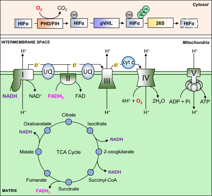
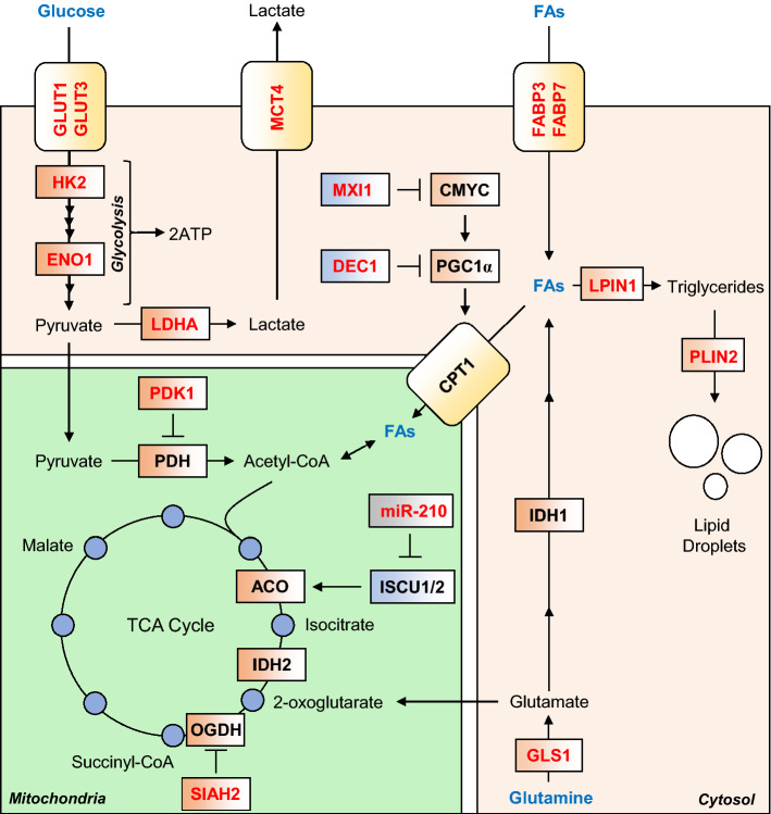
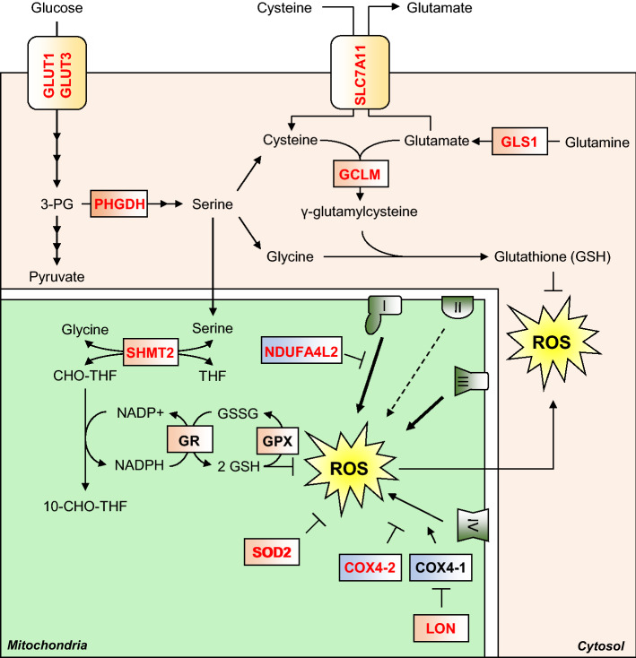
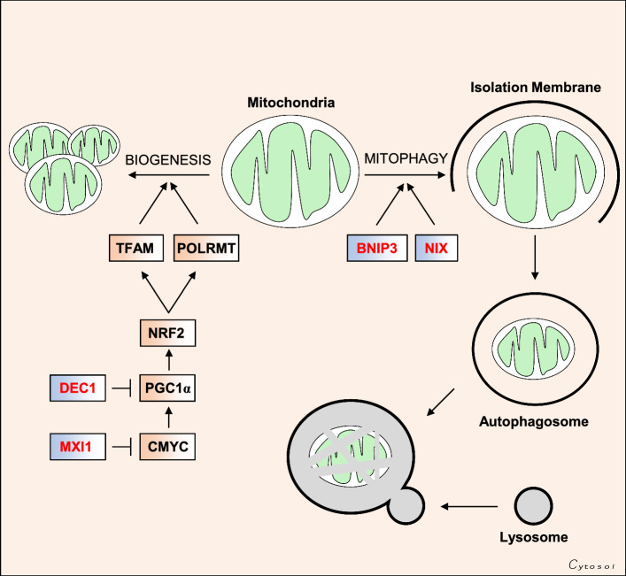
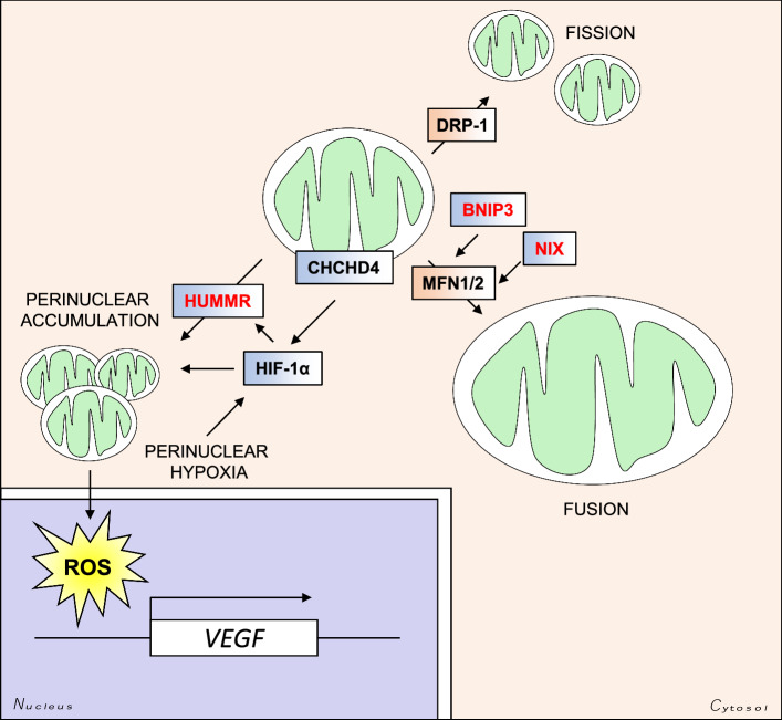
Oxygen is required for the survival of the majority of eukaryotic organisms, as it is important for many cellular processes. Eukaryotic cells utilize oxygen for the production of biochemical energy in the form of adenosine triphosphate (ATP) generated from the catabolism of carbon-rich fuels such as glucose, lipids and glutamine. The intracellular sites of oxygen consumption-coupled ATP production are the mitochondria, double-membraned organelles that provide a dynamic and multifaceted role in cell signalling and metabolism. Highly evolutionarily conserved molecular mechanisms exist to sense and respond to changes in cellular oxygen levels. The primary transcriptional regulators of the response to decreased oxygen levels (hypoxia) are the hypoxia-inducible factors (HIFs), which play important roles in both physiological and pathophysiological contexts. In this review we explore the relationship between HIF-regulated signalling pathways and the mitochondria, including the regulation of mitochondrial metabolism, biogenesis and distribution.

Keywords: HIF; Hypoxia; Metabolism; Mitochondrial biogenesis; Oxphos; Oxygen; Respiratory chain.


Figures

Fig. 1
Fig. 1
Oxygen-dependent ATP synthesis and HIF-α degradation. ATP is synthesised in the mitochondria by an F1F0-type ATP synthase, also known as CV (V). It uses energy provided by an electrochemical gradient formed through proton (H +) pumping between the mitochondrial matrix and the intermembrane space, which is carried out by CI, CIII and CIV. Proton transfer against this electrochemical gradient is energetically unfavourable, and is therefore powered by serial transfer of electrons (e–) from CI and CII to CIII via ubiquinone (UQ), and from CIII to CIV via cytochrome c (cyt c). CIV combines these electrons with molecular oxygen (O2) and protons to produce water (H2O). Electrons are provided to the ETC by the reducing equivalents NADH (to CI) and FADH2 (to CII), which are produced at various steps in the TCA cycle. Oxygen is also used to regulate the stability of HIF-α subunits. In the presence of oxygen, PHD and FIH enzymes hydroxylate specific residues on HIF-α proteins, which permit their recognition and ubiquitination by pVHL. Polyubiquitination of HIF-α proteins then targets them for degradation by the 26S proteasome
Fig. 2
Fig. 2
HIF-mediated changes to mitochondrial carbon metabolism. HIF signalling mediates an increase in anaerobic ATP production, by increasing glycolysis rates, through increased expression of glucose transporters GLUT1 and GLUT3, and almost all glycolytic enzymes, such as HK2 and ENO1. HIF signalling also diverts glucose-derived pyruvate away from mitochondrial respiration by increasing expression of LDHA, and PDK1, a negative regulator of PDH. Lactate efflux is increased by HIF-mediated increase in expression of MCT4. Fatty acids (FAs) are also diverted from catabolism to acetyl-CoA, through suppression of the rate-limiting enzyme in the mitochondrial import of FAs, CPT1. This is achieved through HIF-dependent upregulation of two negative regulators of PGC-1α expression, namely MXI1 and DEC1. FA import into the cell is increased by HIF-dependent upregulation of FABP3 and FABP7, and their conversion to triglycerides and lipid droplets is increased, through upregulation of LPIN1 and PLIN2, respectively. Glutamine is diverted from oxidation by the TCA cycle, through degradation of the 2-OG metabolising enzyme OGDH, by HIF-mediated increase in the expression of the OGDH-targeting SIAH2. This increases 2-OG availability for reductive carboxylation via IDH 1 and IDH2, which produces lipogenic acetyl-CoA. Glutamine flux to acetyl-CoA is also increased by HIF-dependent upregulation of GLS1. Conversely, oxidation of acetyl-CoA to 2-OG is suppressed through HIF-dependent upregulation of mir-210, which downregulates ISCU1/2, which is required for the activity of ACO
Fig. 3
Fig. 3
HIF-mediated suppression of ROS. Hypoxia stimulates ROS production, which can damage macromolecules such as proteins, lipids and DNA. HIF signalling upregulates synthesis of the antioxidant tripeptide glutathione by multiple means. HIF upregulates expression of the rate-limiting enzyme in glutathione (GSH) synthesis, GCLM, as well as proteins which increase the cellular levels of the three constituent amino acids of glutathione. Cysteine import is increased by upregulation of SLC7A11, while glutamate synthesis is increased by upregulation of GLS1. Glycine synthesis is increased by increased serine metabolism, first by its synthesis from glycolysis-derived 3-PG, via HIF-dependent upregulation of PHGDH, and second through its conversion to glycine via upregulation of the folate cycle enzyme SHMT2. The folate cycle also produces NADPH, which is utilised by GR to recycle glutathione disulphide (GSSG) to the ROS scavenging GSH. HIF signalling also upregulates the expression of SOD2, a mitochondrial enzyme capable of converting the superoxide free radical to H2O2. Subunit switching is another way the HIF pathway reduces ROS production is also reduced by HIF-mediated subunit switching of the ETC complexes. The HIFs upregulate expression of an alternative subunit of CI (I), NDUFAL2 which produces less ROS than isoform 1. Similarly, HIFs upregulate an alternative subunit of CIV (IV), COX4-2 which produces less ROS, as well as LON, which degrades isoform 2 (COX4-1)
Fig. 4
Fig. 4
HIF-mediated regulation of mitochondrial mass. The HIF pathway reduces mitochondrial number in the cell by suppressing mitochondrial biogenesis and increasing mitochondrial degradation through mitophagy. Mitochondrial biogenesis is regulated by the PGC-1α pathway, which upregulates mitochondrial proteins required for expression of genes encoded by the mitochondrial genome, such as TFAM and POLRMT. This is achieved through HIF-dependent upregulation of two negative regulators of PGC-1α activity, namely MXI1, which inhibits C-MYC-directed PGC-1α expression, and DEC1, which inhibits PGC-1α transcription by binding to its promoter. HIF signalling also upregulates the expression of two related proteins expressed on the mitochondrial outer membrane, namely BNIP3 and NIX (BNIP3L). These proteins flag mitochondria for degradation by the autophagy pathway
Fig. 5
Fig. 5
HIF-mediated regulation of mitochondrial size and intracellular distribution. HIF targets regulate the subcellular distribution of mitochondria. The HIF-target HUMMR regulates mitochondrial trafficking along microtubules, and promotes perinuclear clustering of the mitochondrial network. The mitochondrial DRS protein CHCHD4 also stimulates perinuclear clustering of the mitochondria in a HIF-dependent manner. Perinuclear accumulation of the mitochondria stimulates ROS-mediated HIF-dependent upregulation of VEGF transcription. The HIF-regulated genes BNIP3 and NIX (BNIP3L) are involved in the MFN1- and MFN2-dependent fusion of mitochondria. Enlargement of the mitochondria confers increased mitochondrial membrane integrity, and protection against apoptosis
Fig. 6
Fig. 6
Mitochondrial regulation of HIF signalling. Mitochondrial oxygen consumption at CIV (IV) regulates the intracellular availability of oxygen, which is required for hydroxylation of HIF-α subunits by the PHD enzymes. The mitochondria also metabolise the PHD substrate 2-OG, and consequently regulate its intracellular levels. Two products of mitochondrial 2-OG metabolism, succinate and fumarate, are capable of competitively inhibiting PHD activity, and thus TCA cycle enzyme activity influences intracellular levels of these metabolites, and influence PHD-mediated HIF-α hydroxylation. Disease-associated mutations in IDH2 cause a neoenzymatic reaction which produces 2-HG which is also a competitive inhibitor of the PHD enzymes. ROS production by the mitochondria also regulates PHD activity by influencing the REDOX state of the ferrous (Fe) ion cofactor

Similar articles

Cited by

References

    1. Rich PR. The molecular machinery of Keilin’s respiratory chain. Biochem Soc Trans. 2003;31:1095–1105. doi: 10.1042/bst0311095. - DOI - PubMed
    1. Wallimann T, Wyss M, Brdiczka D, Nicolay K, Eppenberger HM. Intracellular compartmentation, structure and function of creatine kinase isoenzymes in tissues with high and fluctuating energy demands: the ‘phosphocreatine circuit’ for cellular energy homeostasis. Biochem J. 1992;281(Pt 1):21–40. doi: 10.1042/bj2810021. - DOI - PMC - PubMed
    1. Wyss M, Kaddurah-Daouk R. Creatine and creatinine metabolism. Physiol Rev. 2000;80:1107–1213. doi: 10.1152/physrev.2000.80.3.1107. - DOI - PubMed
    1. Carlile MJ, Watkinson SC, Gooday GW. The fungi. 2. San Diego: Academic Press; 2001.
    1. Hochachka PW, Mustafa T. Invertebrate facultative anaerobiosis. Science. 1972;178:1056–1060. doi: 10.1126/science.178.4065.1056. - DOI - PubMed

MeSH terms

LinkOut - more resources