Review
. 2019 Feb;8(2):10.1128/ecosalplus.ESP-0034-2018.
doi: 10.1128/ecosalplus.ESP-0034-2018.

Architecture, Function, and Substrates of the Type II Secretion System

Affiliations
Review

Architecture, Function, and Substrates of the Type II Secretion System

Konstantin V Korotkov et al. EcoSal Plus. 2019 Feb.

Abstract

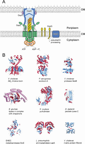
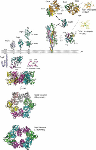
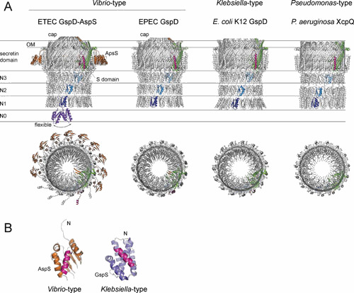
The type II secretion system (T2SS) delivers toxins and a range of hydrolytic enzymes, including proteases, lipases, and carbohydrate-active enzymes, to the cell surface or extracellular space of Gram-negative bacteria. Its contribution to survival of both extracellular and intracellular pathogens as well as environmental species of proteobacteria is evident. This dynamic, multicomponent machinery spans the entire cell envelope and consists of a cytoplasmic ATPase, several inner membrane proteins, a periplasmic pseudopilus, and a secretin pore embedded in the outer membrane. Despite the trans-envelope configuration of the T2S nanomachine, proteins to be secreted engage with the system first once they enter the periplasmic compartment via the Sec or TAT export system. Thus, the T2SS is specifically dedicated to their outer membrane translocation. The many sequence and structural similarities between the T2SS and type IV pili suggest a common origin and argue for a pilus-mediated mechanism of secretion. This minireview describes the structures, functions, and interactions of the individual T2SS components and the general architecture of the assembled T2SS machinery and briefly summarizes the transport and function of a growing list of T2SS exoproteins. Recent advances in cryo-electron microscopy, which have led to an increased understanding of the structure-function relationship of the secretin channel and the pseudopilus, are emphasized.


Figures

Figure 1
Figure 1
Overview of the general architecture of the T2SS and its substrates. (A) A schematic diagram of topology and location of the conserved core components of the T2SS. The accessory components GspN, GspA, and GspB are not shown. (B) A selection of the T2SS substrates of variable functions. Protein toxins include V. cholerae AB5 cholera toxin (139) and P. aeruginosa exotoxin A (140). Hydrolytic enzymes include V. cholerae VesB (68), B. glumae lipase in complex with chaperone (shown in purple) (71), K. oxytoca pullulanase (77), D. dadantii pectate lyase C (141), EHEC metalloprotease StcE (142), and L. pneumophila aminopeptidase LapA (91). V. cholerae biofilm matrix protein RbmA is a scaffolding protein (143, 144).
Figure 2
Figure 2
Structures of the T2SS assembly platform and pseudopilus components. The ATPase is hexameric V. cholerae GspE with C6 and C2 symmetries (20). A close-up view shows the Zn2+ binding site, which is required for the function of GspE (14, 145). Inner membrane components include the cytoplasmic domain of V. cholerae GspF (19), cytoplasmic domain of GspL in complex with N1 domain of V. cholerae GspE (16), periplasmic domain of V. parahaemolyticus GspL (26), periplasmic domain of V. cholerae GspM (25), the homology region (HR) domain of ETEC GspC (32), and the PDZ domain of V. cholerae GspC (29). The structure of periplasmic domain of P. aeruginosa GspL (XcpY) has been recently published (146). Regarding pseudopilus components, in the K. oxytoca GspG pseudopilus model based on the cryo-EM reconstruction (50), a close-up view shows the Ca2+ binding site of K. oxytoca GspG, V. cholerae minor pseudopilin GspH (47), and the trimeric complex of ETEC GspK-GspI-GspJ (48), and a close-up view shows a double-Ca2+ binding site of GspK. The structure of a homologous XcpX-XcpV-XcpW complex from P. aeruginosa has been recently reported (147).
Figure 3
Figure 3
Structures of the T2SS secretins and pilotins. (A) The side and top views of ETEC GspD-AspS complex (37), EPEC GspD (36), E. coli K-12 GspD (34), and P. aeruginosa GspD (35). A single secretin protomer is highlighted, with N1, N2, and N3 domains in shades of blue, the secretin domain in green, and the S domain in magenta. Several AspS protomers (brown) were omitted to clearly show the location of the S domain. The cap subdomain in the Vibrio-type secretins is highlighted in orange. The N0 domains (purple) were not resolved in the available cryo-EM reconstructions due to flexibility. Instead, its approximate location is indicated (148). Note that the N1-N2 domains of EPEC GspD (36) and the N1 domain of P. aeruginosa GspD (35) have been placed as rigid fit models. (B) Structures of pilotins in complex with the secretin S domains (magenta). Structures of Vibrio-type ETEC AspS (37) and Klebsiella-type D. dadantii GspS (116) are shown.

References

    1. Abby SS, Cury J, Guglielmini J, Néron B, Touchon M, Rocha EP. 2016. Identification of protein secretion systems in bacterial genomes. Sci Rep 6:23080. 10.1038/srep23080. - DOI - PMC - PubMed
    1. Cianciotto NP, White RC. 2017. Expanding role of type II secretion in bacterial pathogenesis and beyond. Infect Immun 85:e00014-17. 10.1128/IAI.00014-17. - DOI - PMC - PubMed
    1. Korotkov KV, Sandkvist M, Hol WGJ. 2012. The type II secretion system: biogenesis, molecular architecture and mechanism. Nat Rev Microbiol 10:336–351. 10.1038/nrmicro2762. - DOI - PMC - PubMed
    1. Sikora AE. 2013. Proteins secreted via the type II secretion system: smart strategies of Vibrio cholerae to maintain fitness in different ecological niches. PLoS Pathog 9:e1003126. 10.1371/journal.ppat.1003126. - DOI - PMC - PubMed
    1. Liles MR, Edelstein PH, Cianciotto NP. 1999. The prepilin peptidase is required for protein secretion by and the virulence of the intracellular pathogen Legionella pneumophila. Mol Microbiol 31:959–970. 10.1046/j.1365-2958.1999.01239.x. - DOI - PubMed

Publication types

MeSH terms

LinkOut - more resources