Review
. 2019 Feb 14;8(2):160.
doi: 10.3390/cells8020160.

Cilia Distal Domain: Diversity in Evolutionarily Conserved Structures

Affiliations
Review

Cilia Distal Domain: Diversity in Evolutionarily Conserved Structures

Helena Soares et al. Cells. .

Abstract

Eukaryotic cilia are microtubule-based organelles that protrude from the cell surface to fulfill sensory and motility functions. Their basic structure consists of an axoneme templated by a centriole/basal body. Striking differences in ciliary ultra-structures can be found at the ciliary base, the axoneme and the tip, not only throughout the eukaryotic tree of life, but within a single organism. Defects in cilia biogenesis and function are at the origin of human ciliopathies. This structural/functional diversity and its relationship with the etiology of these diseases is poorly understood. Some of the important events in cilia function occur at their distal domain, including cilia assembly/disassembly, IFT (intraflagellar transport) complexes' remodeling, and signal detection/transduction. How axonemal microtubules end at this domain varies with distinct cilia types, originating different tip architectures. Additionally, they show a high degree of dynamic behavior and are able to respond to different stimuli. The existence of microtubule-capping structures (caps) in certain types of cilia contributes to this diversity. It has been proposed that caps play a role in axoneme length control and stabilization, but their roles are still poorly understood. Here, we review the current knowledge on cilia structure diversity with a focus on the cilia distal domain and caps and discuss how they affect cilia structure and function.

Keywords: cilia; cilia distal domain; cilia structural diversity; microtubule-capping structures.


Conflict of interest statement

The authors declare no conflicts of interest.

Figures
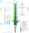
Figure 1
Figure 1
Structural comparison between motile and primary cilia. The cilia axoneme is composed of nine pairs of microtubules (green tubules) that extend from a modified centriole named basal body. Three structural and functional cilia domains can be identified, namely the distal, the middle and the proximal domains, according to their position in relation to the basal body. Cross-section 1 shows the three most common axonemal architectures: (9+0) immotile and motile cilia, with nine microtubule doublets arranged radially; (9+2) motile cilia, with nine microtubule doublets and a central pair of microtubule singlets. Motor proteins (outer dynein arms (yellow) and inner dynein arms (blue)) together with radial spokes generate the wave-like beating of the (9+2) motile cilia; (9+0) motile cilia, with nine microtubule doublets and without central pair of microtubule singlets. In (9+0) motile cilia, motor proteins (outer dynein arms (yellow) and inner dynein arms (blue)) generate a rotational movement. Cross-sections 2 and 3 respectively show, the Y-shaped links (yellow) of the transition zone and the transition fibers (light blue), that link to the membrane creating the ciliary gate. The rootlet (grey) extends from the base of the basal body towards the nucleus and appears as long striated fibers. The basal foot (motile cilia)/basal feet (immotile cilia) (dark blue) provide additional support to the cilia. In the case of motile cilia, the basal foot aligns with the cilia beating direction.
Figure 2
Figure 2
Comparison of the Structural Organization and major components of basal feet from primary and motile cilia. Primary and motile cilia basal feet present different components and domain organizations as observed by super-resolution microscopy. Primary cilia have multiple basal feet whose components are organized in three distinct domains. The most distal region (III) is characterized by the presence of Ninein (NIN) and Centrosomal protein of 170 kDa (CEP170), proteins involved in microtubule (MT) anchoring and components of the centriole sub-distal appendages. The next region (II) contains proteins that are possibly involved in the scaffolding of the basal feet, namely Outer dense fiber protein 2 (ODF2). The most proximal region (I) to the basal body was named “basal body anchoring” although there was no protein assigned to this region. Motile cilia present only one basal foot, aligned with the cilia beating direction. The domain organization is simpler than that of primary cilia, corresponding, to a certain extent to a rearrangement of Regions II and III. On the most distal region of the basal foot (II), besides the proteins in common with the basal feet of primary cilia (i.e. NIN, CEP170, Centriolin (CNTRL)), there are proteins of the γ-tubulin ring complex (γ-TURC) microtubule nucleating complex (i.e. γ-tubulin and protein NEDD1). The most proximal region (I) contains proteins that are common to the primary cilia basal feet (i.e. Centrosomal protein of 128 kDa (CEP128) and ODF2) that are most likely responsible for the scaffolding (based on proteomics and super-resolution microscopy data in [53]).
Figure 3
Figure 3
Diversity of cilia distal domain architectures. (a) Schematic representation of representative cilia tip structures. (I) In trachea cilia, the axoneme structure is (9+2) and at the tip the A-tubules and central pair are bound to a central cap that is attached to the ciliary crown, a structure constituted by a cluster of fibrils that project from the membrane to the outside of the cilia tip. (II) In the ciliate protozoa (Tetrahymena) and green alga (Chlamydomonas), the axoneme structure is (9+2). At the tip, the A-tubules are attached to the membrane by distal filaments that, in their proximal ends, form a plug which inserts into A-tubule lumen, while the central microtubule pair is bound to the central cap. (III). In the parasite protozoa Leishmania mexicana amastigote, the axoneme structure is (9+0). Distally, the 9-fold symmetry is broken by microtubule doublets progressively occupying a more central position. (IV) In the flat worm turbellarian the cilia present a typical (9+2) axonemal pattern through the main part of its length. Near the tip, this pattern changes to one where the microtubule central pair and doublets 1, 2, 3, 8 and 9 terminate in a distal cap that is attached to a ciliary crown, whereas doublets 4 to 7 end at a dense material of a proximal secondary cap. Therefore, the tip is asymmetric. (V) In the kidney primary cilia, the axoneme is (9+0) and progressively, along the axoneme, microtubule doublets suffer a displacement towards the center and the axoneme narrows towards the tip. In the distal segment, doublets are converted into singlets through the loss of the B-tubule. (VI). In the parasite protozoa Trypanosome brucei, the tip is (9+2) but the microtubule central pair is slightly shorter. Microtubule doublets and central pair present a dense material at the ends. (VII) In the adult Caenorhabditis elegans sensory amphid channel cilia (yellow) the axoneme is (9+a few singlets), and the microtubules maintain a radial organization and in vertebrate photoreceptors (purple) the axoneme is (9+0). However, both tips present only microtubule singlets. The differences can be observed at the cross section (VIII). In the adult Caenorhabditis elegans’ sensory amphid wing cilia C, the middle region of the axoneme contains both doublets and singlets that “splay apart” laterally and end together ~0.5 μm below the distal membrane. The tip presents a membranous fan-like structure. (b) Macrocilia of the ctenophore Beroe. Macrocilia present hundreds of ciliary axonemes with a (9+2) structure surrounded by a common membrane, with a giant capping structure. At the tip, doublet microtubules end and only singlet microtubules remain, linked together by electron-dense material. Central microtubule pairs are often still recognizable at this level. Schemes were based on: [58,92,103,109,110,111,112].
Figure 4
Figure 4
Several stages of Tetrahymena cilia assembly model. Axoneme assembly: (A) Three-fold structures present at the transition region and axonemes (210–460 nm) of growing Tetrahymena cilia. It was speculated that the three-fold structures function like ring sections that will progressively assemble transversally, originating a full ring (9-fold structure) contributing to the axonemal growth. Cap assembly: (A) A collar-like shape is assembled in the axonemal distal tip, shown in red, providing an incomplete cap to the cilium. The other components of the cap are larger structures that are visualized near the edge of the cilium tip (A); (B) All cap components are assembled and finally originate the full mature cap with a dome shape. In blue, the axoneme wall; in transparent dark blue the three-fold structures. The different components involved in cap assembly are shown in red. The AFM topographic images representative of the different steps of cilia and cap assembly are shown at the top of the schematic drawing. Reprinted with permission from Reference [150].
Figure 5
Figure 5
The dynamic distal domain and associated proteins. Important events in cilia function occur at their distal domain like cilia assembly/disassembly, intraflagellar transport (IFT) complexes’ remodeling, and signal reception/transduction. The IFT system forms cargo-carrying trains moved along the axoneme through the action of kinesin-2 (anterograde) and dynein-2 (retrograde) motors. This transport is essential for the assembly and maintenance of cilia and flagella. Tubulin subunits are transported throughout the cilium by IFT. At the tip, IFT trains split apart and mix with each other to assemble into new trains, which move back to the cell body with new cargos. Kinesin transports dynein to the tip as inactive cargo, disconnects from IFT trains at the tip and turns around by diffusion. Distinct functional arrays of ciliary proteins that concentrate at the tip are indicated (Molecular chaperones, Microtubule dynamic regulators, Channel proteins and signaling transduction, Cap structural proteins (when cap structures are present), Post-translational modifications enzymes, Actin binding proteins and motors, Anchoring junctions and others). A-tubule singlets are in general characteristic of the distal domain and present a dynamic behavior by polymerization and depolymerization, therefore changing the tip segment length in response to different signals.

Similar articles

Cited by

References

    1. Waters A.M., Beales P.L. Ciliopathies: An expanding disease spectrum. Pediatr. Nephrol. 2011;26:1039–1056. doi: 10.1007/s00467-010-1731-7. - DOI - PMC - PubMed
    1. Ostrowski L.E., Blackburn K., Radde K.M., Moyer M.B., Schlatzer D.M., Moseley A., Boucher R.C. A proteomic analysis of human cilia: Identification of novel components. Mol. Cell Proteom. 2002;1:451–465. doi: 10.1074/mcp.M200037-MCP200. - DOI - PubMed
    1. Haimo L.T., Rosenbaum J.L. Cilia, flagella, and microtubules. J. Cell Biol. 1981;91:125s–130s. doi: 10.1083/jcb.91.3.125s. - DOI - PMC - PubMed
    1. Dellinger O.P. The cilium as a key to the structure of contractile protoplasm. J. Morphol. 1909;20:171–210. doi: 10.1002/jmor.1050200202. - DOI
    1. Schmitt F.O., Hall C.E., Jakus M.A. The Ultrastructure of Protoplasmic Fibrils. Biol. Symp. 1943;10:261–276.

Publication types

LinkOut - more resources