. 2019 Feb 14;11(2):404.
doi: 10.3390/nu11020404.

HM-Chromanone Isolated from Portulaca oleracea L. Protects INS-1 Pancreatic β Cells against Glucotoxicity-Induced Apoptosis

Affiliations

HM-Chromanone Isolated from Portulaca oleracea L. Protects INS-1 Pancreatic β Cells against Glucotoxicity-Induced Apoptosis

Jae Eun Park et al. Nutrients. .

Abstract

In this study, we investigated whether (E)-5-hydroxy-7-methoxy-3-(2'-hydroxybenzyl)-4-chromanone, a homoisoflavonoid compound isolated from Portulaca oleracea L., protects INS-1 pancreatic β cells against glucotoxicity-induced apoptosis. Treatment with high glucose (30 mM) induced apoptosis in INS-1 pancreatic β cells; however, the level of cell viability was significantly increased by treatment with (E)-5-hydroxy-7-methoxy-3-(2'-hydroxybenzyl)-4-chromanone. Treatment with 10⁻20 µM of (E)-5-hydroxy-7-methoxy-3-(2'-hydroxybenzyl)-4-chromanone dose-dependently increased cell viability and significantly decreased the intracellular level of reactive oxygen species (ROS), thiobarbituric acid reactive substances (TBARS), and nitric oxide levels in INS-1 pancreatic β cells pretreated with high glucose. These effects were associated with increased anti-apoptotic Bcl-2 protein expression, while reducing pro-apoptotic Bax, cytochrome C, and caspase 9 protein expression. Treatment with (E)-5-hydroxy-7-methoxy-3-(2'-hydroxybenzyl)-4-chromanone reduced the apoptosis previously induced by high-level glucose-treatment, according to annexin V/propidium iodide staining. These results demonstrate that (E)-5-hydroxy-7-methoxy-3-(2'-hydroxybenzyl)-4-chromanone may be useful as a potential therapeutic agent to protect INS-1 pancreatic β cells against high glucose-induced apoptosis.

Keywords: (E)-5-hydroxy-7-methoxy-3-(2′-hydroxybenzyl)-4-chromanone; INS-1 pancreatic β cell; Portulaca oleracea; glucotoxicity.


Conflict of interest statement

The authors declare no conflict of interest.

Figures
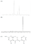
Figure 1
Figure 1
1H NMR, 13C NMR, and chemical structure of HM-Chromanone isolated from P. oleracea. 1H NMR, 13C NMR, and chemical structure of HM-Chromanone isolated from P. oleracea. (A) 1H NMR. (B) 13C NMR spectrum, and (C) chemical structure of (E)-5-hydroxy-7-methoxy-3-(2′-hydroxybenzyl)-4-chromanone (HM-chromanone).
Figure 2
Figure 2
Effect of HM-chromanone on cell viability in high glucose-treated INS-1 pancreatic β cells. INS-1 pancreatic β cells (2 × 10⁴ cells/well) were preincubated in 96-well plates with 5.5 or 30 mM glucose for 48 h, and then incubated with HM-chromanone (0, 1, 5, 10, or 20 µM) for 48 h. The 5.5 mM glucose represents normal glucose, while the 30 mM glucose represents high glucose concentrations. Each value is expressed as the mean ± standard deviation (SD) (n = 3). a~e Values with different letters were significantly different at p < 0.05, as analyzed by Duncan’s multiple-range test.
Figure 3
Figure 3
Effect of HM-chromanone on intracellular levels of reactive oxygen species (ROS) in high glucose-treated INS-1 pancreatic β cells. INS-1 pancreatic β cells (2 × 10⁴ cells/well) were preincubated with 5.5 or 30 mM glucose in 96-well plates for 48 h, and then incubated with HM-chromanone (0, 1, 5, 10, or 20 µM) for 48 h. The concentration of 5.5 mM glucose represents normal glucose, while the 30 mM glucose represents a high glucose concentration. Each value is expressed as the mean ± standard deviation (n = 3). a~f Values with different letters were significantly different at p < 0.05, as analyzed by Duncan’s multiple-range test.
Figure 4
Figure 4
Effect of HM-chromanone on the generation of thiobarbituric acid reactive substances (TBARS) in high glucose-treated INS-1 pancreatic β cells. INS-1 pancreatic β cells (2 × 10⁴ cells/well) were preincubated in 96-well plates with 5.5 or 30 mM glucose for 48 h, and then incubated with HM-chromanone (0, 1, 5, 10, or 20 µM) for 48 h. The concentration of 5.5 mM glucose represents normal glucose, while 30 mM glucose represents a high glucose concentration. Each value is expressed as the mean ± standard deviation (n = 3). a~f Values with different letters were significantly different at p < 0.05, as analyzed by Duncan’s multiple-range test.
Figure 5
Figure 5
Effect of HM-chromanone on the level of nitric oxide (NO) in high glucose-treated INS-1 pancreatic β cells. INS-1 pancreatic β cells (2 × 10⁴ cells/well) were preincubated in 96-well plates with 5.5 or 30 mM glucose for 48 h, and then incubated with HM-chromanone (0, 1, 5, 10, or 20 µM) for 48 h. The concentration of 5.5 mM glucose represents normal glucose, while 30 mM glucose represents a high glucose concentration. Each value is expressed as the mean ± standard deviation (n = 3). a~f Values with different letters were significantly different at p < 0.05, as analyzed by Duncan’s multiple-range test.
Figure 6
Figure 6
Effect of HM-chromanone on insulin secretion in high glucose-treated INS-1 pancreatic β cells. INS-1 pancreatic β cells (2 × 10⁵ cells/well) were preincubated in 96-well plates with 5.5 or 30 mM glucose, and then incubated with HM-chromanone (0, 1, 5, 10, or 20 µM) for 48 h. Thereafter, the cells were stimulated with Krebs–Ringer buffer containing 5 or 25 mM glucose for 60 min. The 5.5 mM concentration of glucose represents normal glucose, while 30 mM glucose represents a high glucose concentration. Each value is expressed as the mean ± standard deviation (n = 3). a~f Values with different letters were significantly different at p < 0.05, as analyzed by Duncan’s multiple-range test.
Figure 7
Figure 7
Effect of HM-chromanone on expressions of Bax, Bcl-2, cytochrome C, caspase 9, and caspase 3 in high glucose-treated INS-1 pancreatic β cells. INS-1 pancreatic β cells were preincubated with 5.5 or 30 mM glucose for 48 h, and then incubated with HM-chromanone (10 or 20 µM) for 48 h. Equal amounts of cell lysates were electrophoresed and levels of Bax, Bcl-2, cytochrome C, caspase 9, and caspase 3 protein expression were measured using western blots. (A) Bax, Bcl-2, cytochrome C, caspase 9, and caspase 3 protein levels. (B) Expression levels of Bax, Bcl-2, cytochrome C, caspase 9, and caspase 3. Each value is expressed as the mean ± standard deviation (n = 3). a~d Values with different letters were significantly different at p < 0.05, as analyzed by Duncan’s multiple-range test.
Figure 8
Figure 8
Identification of the type of cell death by Annexin V-FITC/propidium iodide (PI) staining. The status of apoptotic cell death was determined by counting INS-1 pancreatic β cells stained with annexin V-FITC/PI using a flow cytometer. Cells were preincubated with glucose and then incubated in the presence or absence of HM-chromanone (10 or 20 µM). The lower and upper right quadrants show the numbers of early and late apoptotic cells, respectively.

References

    1. Butler A.E., Janson J., Bonner-Weir S., Ritzel R., Rizza R.A., Butler P.C. Beta-cell deficit and increased beta-cell apoptosis in humans with type 2 diabetes. Diabetes. 2003;52:102–110. doi: 10.2337/diabetes.52.1.102. - DOI - PubMed
    1. Kahn S.E. The relative contributions of insulin resistance and beta-cell dysfunction to the pathophysiology of Type 2 diabetes. Diabetologia. 2003;46:3–19. doi: 10.1007/s00125-002-1009-0. - DOI - PubMed
    1. UK Prospective Diabetes Study (UKPDS) Group Intensive blood-glucose control with sulphonylureas or insulin compared with conventional treatment and risk of complications in patients with type 2 diabetes (UKPDS 33) Lancet. 1998;352:837–853. doi: 10.1016/S0140-6736(98)07019-6. - DOI - PubMed
    1. Rashid M.A., Lee S., Tak E. Carbonyl reductase 1 protects pancreatic β-cells against oxidative stress-induced apoptosis in glucotoxicity and glucolipotoxicity. Radic. Biol. Med. 2010;49:1522–1533. doi: 10.1016/j.freeradbiomed.2010.08.015. - DOI - PubMed
    1. Sung Y.A., Hong Y.S. Mechanism of the insulin secretory defect by chronically elevated glucose levels in pancreatic islets: Depletion of insulin content due to hyperstimulation by glucose. J. Kor. Diabetes Asso. 2000;24:1–9.

MeSH terms