. 2019 Feb 15;11(2):158.
doi: 10.3390/v11020158.

THO Complex Subunit 7 Homolog Negatively Regulates Cellular Antiviral Response against RNA Viruses by Targeting TBK1

Affiliations

THO Complex Subunit 7 Homolog Negatively Regulates Cellular Antiviral Response against RNA Viruses by Targeting TBK1

Tian-Sheng He et al. Viruses. .

Abstract

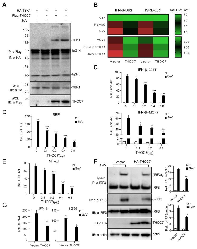
RNA virus invasion induces a cytosolic RIG-I-like receptor (RLR) signaling pathway by promoting assembly of the Mitochondrial antiviral-signaling protein (MAVS) signalosome and triggers the rapid production of type I interferons (IFNs) and proinflammatory cytokines. During this process, the pivotal kinase TANK binding kinase 1 (TBK1) is recruited to the MAVS signalosome to transduce a robust innate antiviral immune response by phosphorylating transcription factors interferon regulatory factor 3 (IRF3) and nuclear factor (NF)-κB and promoting their nuclear translocation. However, the molecular mechanisms underlying the negative regulation of TBK1 are largely unknown. In the present study, we found that THO complex subunit 7 homolog (THOC7) negatively regulated the cellular antiviral response by promoting the proteasomal degradation of TBK1. THOC7 overexpression potently inhibited Sendai virus- or polyI:C-induced IRF3 dimerization and phosphorylation and IFN-β production. In contrast, THOC7 knockdown had the opposite effects. Moreover, we simulated a node-activated pathway to show that THOC7 regulated the RIG-I-like receptors (RLR)-/MAVS-dependent signaling cascade at the TBK1 level. Furthermore, THOC7 was involved in the MAVS signalosome and promoted TBK1 degradation by increasing its K48 ubiquitin-associated polyubiquitination. Together, these findings suggest that THOC7 negatively regulates type I IFN production by promoting TBK1 proteasomal degradation, thus improving our understanding of innate antiviral immune responses.

Keywords: MAVS signalosome; TBK1; THOC7; cellular antiviral response.


Conflict of interest statement

The authors declare no competing financial interests.

Figures

Figure 1
Figure 1
Overexpression of THOC7 negatively regulates the production of type I interferons (IFNs). (A) Interaction of THOC7 with TBK1 in a mammalian overexpression system. 293T cells were transfected with mock control, Flag-THOC7, and HA-TBK1 (10 μg each), followed by treatment with Sev or not for 10 h. At 24 h post-transfection, co-immunoprecipitation was performed with anti-HA beads and immunoblotting analysis with anti-Flag antibodies. (B) Small-scale screening of THOC7 functions in regulating SeV- or polyI:C-induced type I IFN signaling. 293T cells were seeded into 24-well plates and transfected with a luciferase reporter gene carrying the ISRE or IFN-β promoter (100 ng/well), pRL-TK (50 ng/well) and THOC7, followed by infection with SeV or transfection with polyI:C/TBK1 for 12 h. Relative luciferase activity levels were arbitrarily set to 0.1 (green). (CE) THOC7 inhibited IFN-β promoter, ISRE, and NF-κB luciferase activation in a dose-dependent manner. Similar luciferase assay was performed, except with increasing amounts of the expression vector for THOC7. (F) THOC7 significantly reduced the phosphorylation and dimerization of IRF3. 293T cells were seeded into 6-well plates and transfected with HA-THOC7 (4 μg). After transfection for 12 h, the cells were treated with SeV or not for 10 h. Lysates were analyzed by native PAGE or SDS-PAGE. Quantification of western blotting bands from three independent experiments was performed with Image J software. (G) THOC7 inhibits IFN-β gene and ISG56 transcription. Similar transfection as described for (F) was performed. The mRNA levels were measured by q-PCR. Error bars indicate SD. n = 3. *, p < 0.05; **, p < 0.01; ***, p < 0.001; ns, no significant difference.
Figure 2
Figure 2
Knockdown of THOC7 leads to up-regulation of type I IFNs. (A) Immunoblot analysis following knockdown of transfected THOC7 in 293T cells transfected with THOC7-specific RNAis (2 μg), Flag-THOC7 plasmid (1 μg), and Flag-P50 (0.1 μg, as an internal control protein). (B) Real-time PCR of knockdown of exogenous THOC7 (left panel) or IFN-β mRNA (right panel) in 293T cells transfected with control RNAi or RNAis against THOC7 (4 μg). (C) Native PAGE analysis of IRF3 dimerization in 293T cells transfected with THOC7-specific RNAis (4 μg) and treated with SeV for 12 h. (D) Luciferase assays of 293T cells transfected with ISRE or IFNβ luciferase report plasmid (100 ng/well) with pRL-TK (50 ng/well), as well as THOC7 RNAi or control (0.5 μg/well). (E) IRF3 dimerization analysis of THOC7 RNAi#3 in gradient times of SeV treatment. Similar experiments were performed as (C) except with an increased time of SeV treatment. (F) Effects of knockdown of THOC7 on SeV-induced phosphorylation of IRF3. 293T cells were transfected with control RNAi or THOC7 RNAi#3 and infected with SeV for 10 h, followed by immunoblotting analysis with the indicated antibodies. Quantification of western blotting bands (A,C,E,F) from three independent experiments was performed with Image J software. Error bars indicate SD. n = 3. *, p < 0.05; **, p < 0.01; ***, p < 0.001; ns, no significant difference.
Figure 3
Figure 3
THOC7 regulates the RLR signaling pathway by targeting TBK1. (A) A simulation model of node-activated pathway for the role of THOC7 in regulating the RLR signaling pathway. Downstream signaling was activated by transfection of the activated form of signaling pathway molecules containing RIG-I-N, MAVS, TBK1, IKKε, and IRF3-5D. (B,C) Effects of THOC7 overexpression or THOC7 knockdown on IFNβ or ISRE activation mediated by various activated forms of signaling molecules. 293T cells were seeded into 24-well plates and transfected with IFN-β or ISRE luciferase reporter (100 ng/well), pRL-TK (50 ng/well), THOC7 expression vector (0.5 μg, B), or THOC71 RNAi#3 constructs (0.5 μg, C) together with indicated activated forms of signaling molecules. Cells were harvested and analyzed by dual-luciferase reporter assay. After transfection for 24 h, the relative luciferase activities were normalized based on pRL-TK control activities. Error bars indicate SD. n = 3. *, p < 0.05; **, p < 0.01; ***, p < 0.001; ns, no significant difference.
Figure 4
Figure 4
THOC7 is involved in the (mitochondrial antiviral signaling (MAVS) signalosome and drives proteasomal degradation of TBK1. (A) THOC7 was involved in forming the MAVS signaling complex. 293T cells were seeded into 100-mm dishes and transfected with Flag-tagged THOC7 or empty control plasmids (10 μg) and HA-tagged RLR signaling molecules (10 μg) as indicated. After 12 h of transfection, the cells were treated with or without SeV for 12 h. Co-immunoprecipitation and immunoblot analysis was performed with the indicated antibodies. (B) THOC7 promoted degradation of exogenous and endogenous TBK1 in a dose-dependent manner. 293T cells were seeded into 6-well plates and transfected with increasing amounts of Flag-THOC7 (0, 0.1, 0.2, 0.4, 0.8, 1.2 μg), and HA-TBK1 (2 μg). Twenty-four hours after transfection, immunoblot analysis was performed with the indicated antibodies. (C) MG132 restored the TBK1 protein level in the presence of THOC7. 293T cells were seeded into 6-well plates and transfected with Flag-THOC7 (2 μg) and HA-TBK1 (1.5 μg). MG132 and CHX were added after 6 h of transfection and the cells were harvested for analysis after 24 h. (D,E) THOC7 increased the K48-linked ubiquitination of TBK1. 293T cells were seeded into 100-mm dishes and transfected with the indicated plasmids (8 μg each). Similar co-immunoprecipitation and immunoblotting experiments were performed with the indicated antibodies. Quantification of western blotting bands (A,C,E,F) from three independent experiments was performed with Image J software. Error bars indicate SD. n = 3. *, p < 0.05; **, p < 0.01; ***, p < 0.001; ns, no significant difference.

Similar articles

Cited by

References

    1. Medzhitov R. Recognition of microorganisms and activation of the immune response. Nature. 2007;449:819–826. doi: 10.1038/nature06246. - DOI - PubMed
    1. Benko S., Philpott D.J., Girardin S.E. The microbial and danger signals that activate Nod-like receptors. Cytokine. 2008;43:368–373. doi: 10.1016/j.cyto.2008.07.013. - DOI - PubMed
    1. Akira S., Takeda K. Toll-like receptor signalling. Nat. Rev. Immunol. 2004;4:499–511. doi: 10.1038/nri1391. - DOI - PubMed
    1. Takeuchi O., Akira S. Innate immunity to virus infection. Immunol. Rev. 2009;227:75–86. doi: 10.1111/j.1600-065X.2008.00737.x. - DOI - PMC - PubMed
    1. Fritz J.H., Ferrero R.L., Philpott D.J., Girardin S.E. Nod-like proteins in immunity, inflammation and disease. Nat. Immunol. 2006;7:1250–1257. doi: 10.1038/ni1412. - DOI - PubMed

Publication types

MeSH terms

LinkOut - more resources