Comparative Study
. 2019 Feb 15;11(2):159.
doi: 10.3390/v11020159.

Induction of Tier 1 HIV Neutralizing Antibodies by Envelope Trimers Incorporated into a Replication Competent Vesicular Stomatitis Virus Vector

Affiliations
Comparative Study

Induction of Tier 1 HIV Neutralizing Antibodies by Envelope Trimers Incorporated into a Replication Competent Vesicular Stomatitis Virus Vector

C Anika Bresk et al. Viruses. .

Abstract

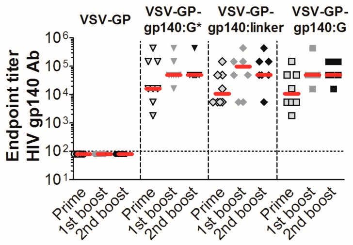
A chimeric vesicular stomatitis virus with the glycoprotein of the lymphocytic choriomeningitis virus, VSV-GP, is a potent viral vaccine vector that overcomes several of the limitations of wild-type VSV. Here, we evaluated the potential of VSV-GP as an HIV vaccine vector. We introduced genes for different variants of the HIV-1 envelope protein Env, i.e., secreted or membrane-anchored, intact or mutated furin cleavage site or different C-termini, into the genome of VSV-GP. We found that the addition of the Env antigen did not attenuate VSV-GP replication. All HIV-1 Env variants were expressed in VSV-GP infected cells and some were incorporated very efficiently into VSV-GP particles. Crucial epitopes for binding of broadly neutralizing antibodies against HIV-1 such as MPER (membrane-proximal external region), CD4 binding site, V1V2 and V3 loop were present on the surface of VSV-GP-Env particles. Binding of quaternary antibodies indicated a trimeric structure of VSV-GP incorporated Env. We detected high HIV-1 antibody titers in mice and showed that vectors expressing membrane-anchored Env elicited higher antibody titers than vectors that secreted Envs. In rabbits, Tier 1A HIV-1 neutralizing antibodies were detectable after prime immunization and titers further increased after boosting with a second immunization. Taken together, VSV-GP-Env is a promising vector vaccine against HIV-1 infection since this vector permits incorporation of native monomeric and/or trimeric HIV-1 Env into a viral membrane.

Keywords: 1086.C HIV-1 Env; HIV vaccine; VSV-GP viral vaccine vector; broadly neutralizing antibodies; vesicular stomatitis virus.


Conflict of interest statement

D.v.L. is an inventor of VSV-GP and held minority shares in the biotech company ViraTherapeutics GmbH, which holds the intellectual property rights for VSV-GP. For the other authors, no competing financial interests exist. The funders had no role in the design of the study; in the collection, analyses, or interpretation of data; in the writing of the manuscript, or in the decision to publish the results.

Figures

Figure 1
Figure 1
Schematic overview of human immunodeficiency virus (HIV) Env variants. The linear structure of HIV gp160 (A) and the three-dimensional structure of the monomers and trimers on the surface of infected cells or viral particles (B) are shown. (C) Schematic overview of HIV Env variants that were cloned into position 5 of VSV-GP vectors. Color coding: gp120 (red), extracellular part of gp41 (green), MPER (membrane-proximal external region; purple), transmembrane domain (TM; solid line for HIV-TM or dotted line for vesicular stomatitis virus glycoprotein (VSV-G)-TM, yellow) and C-terminus (solid line for HIV C-terminus or dotted line for VSV-G C-terminus, dark blue). Constructs contained either a functional furin cleavage site, a mutated furin cleavage site (indicated by an asterisk) or a flexible linker replacing the furin cleavage site (light blue). (i) gp160* = full-length HIV Env with a mutated furin cleavage site, (ii) gp140:G* = gp140 fused to the transmembrane domain and cytoplasmic tail of VSV-G with a mutated furin cleavage site, (iii) gp140:G = gp140 fused to the transmembrane domain and cytoplasmic tail of VSV-G with an intact furin cleavage site, (iv) gp140:G-linker = gp140 fused to the transmembrane domain and cytoplasmic tail of VSV-G with a flexible linker replacing the furin cleavage site, (v) gp120, (vi) Δ147 = truncated HIV Env with only 3 intracellular amino acids at the C-terminus, and an intact furin cleavage site, (vii) TM1 = HIV Env with an SIV segment insertion at the C-terminus, a Y712I mutation, a STOP codon after F717, and a native furin cleavage site, and (viii) TM2 = HIV Env with the SIV segment insertion, a deletion of GY711-712, R722G and S727P mutations (SIVmac239 numbering), a STOP codon after F717, and an intact furin cleavage site. (D) Furin cleavage site configuration and C-terminus for all constructs are depicted.
Figure 2
Figure 2
Incorporation of HIV Env into VSV-GP particles improved antibody responses. (A) BHK-21 cells were infected with VSV-GP, VSV-GP-gp120, VSV-GP-gp160* or VSV-GP-gp140:G* at an multiplicity of infection (MOI) of 0.1. 24 h post-infection, cell lysates were prepared. (B) VSV-GP, VSV-GP-gp120, VSV-GP-gp160* or VSV-GP-gp140:G* were produced and concentrated via a sucrose cushion. Cell lysates (A) or viral particles (B) were analyzed via western blotting with a gp120-specific antibody (16H3). As a loading control, actin- or VSV-N-specific antibodies were used. (C) 293T cells (Mock), 293T cells expressing VSV-G or 293T cells expressing lymphocytic choriomeningitis virus glycoprotein (LCMV-GP) were infected in duplicates with the replication-defective VSV*ΔG-gp140:G* at an MOI of 0.2. The supernatant was collected 24 h later and viruses were concentrated via low-speed overnight centrifugation through a sucrose cushion. Western blotting was performed with purified viruses using an anti-HIV-1 gp120 monoclonal antibody (16H3) to detect HIV Env. A monoclonal antibody against VSV-N was used as loading control. (D) BHK-21 cells were infected with VSV-GP, VSV-GP-gp120, VSV-GP-gp160* or VSV-GP-gp140:G* at an MOI of 0.1 in duplicates. Four, 8, 24, and 48 h after infection, the supernatant was collected and analyzed for viral titer via TCID50 (50% tissue culture infective dose) assay. Mean ± SEM are shown. The dotted line shows the detection limit of the assay. (E) C57BL/6 mice (n = 8) were immunized intramuscularly with 107 TCID50 of VSV-GP, VSV-GP-gp120, VSV-GP-gp160* or VSV-GP-gp140:G* at weeks 0, 4, and 8. Four weeks after each immunization, plasma was collected and analyzed for the titer of gp140-binding antibodies via ELISA. The median for each group (red line) and the titer for each individual animal is shown. The dotted line shows the detection limit of the assay.
Figure 3
Figure 3
All HIV Env variants are expressed at comparable levels in infected BHK-21 cells. BHK-21 cells were infected with VSV-GP, VSV-GP-gp140:G*, VSV-GP-gp160*, VSV-GP-gp140:G-linker, VSV-GP-gp140:G, VSV-GP-Δ147, VSV-GP-TM1 and VSV-GP-TM2 with a MOI of 0.1. Twenty-four hours after infection, cell lysates were prepared for western blotting (A) or cells were trypsinized and analyzed by flow cytometry (B). As a negative control, cell lysates from non-infected BHK-21 cells (Mock) were used. (A) The non-cleaved HIV Env precursor protein (gp160) and the cleaved gp41 protein were detected with an anti-HIV-1 gp41 monoclonal antibody (4E10, upper two blots). The non-cleaved Env precursor and the cleaved gp120 subunit were detected on a separate membrane using an anti-HIV-1 gp120 monoclonal antibody (16H3, third blot). As a loading control, an anti-actin antibody was used (lower blot). (B) Cells were stained with HIV bnAbs against the V1V2 loop of gp120 (PG9, PG16), the V3 loop of gp120 (PGT121), the CD4 binding site (b12, VRC01, 3BNC117, CH106) or the MPER of gp41 (4E10) and fluorescently-labeled anti-human secondary antibodies. As a loading control, the LCMV-GP-specific WEN4 was used. The ratio of the geometric mean fluorescence of every single HIV-specific antibody relative to the geometric mean fluorescence of the LCMV-specific WEN4 was calculated. The graph shows the mean ratio of duplicate samples for each antibody and virus.
Figure 4
Figure 4
HIV Env is incorporated into VSV-GP particles and displays epitopes crucial for binding of bnAbs. The different VSV-GP particles (VSV-GP, VSV-GP-gp140:G*, VSV-GP-gp160*, VSV-GP-gp140:G-linker, VSV-GP-gp140:G, VSV-GP-Δ147, VSV-GP-TM1 and VSV-GP-TM2) were produced on BHK-21 cells and concentrated via low-speed centrifugation using a sucrose cushion. (A) Purified particles were diluted in PBS to a concentration of 109 TCID50/mL and used for western blot analysis. The non-cleaved HIV Env precursor protein gp160 and the cleaved gp41 protein were detected by using an anti-HIV-1 gp41 monoclonal antibody (4E10, upper two blots). The non-cleaved Env precursor gp160 and the cleaved gp120 subunit were detected on a second membrane using an anti-HIV-1 gp120 monoclonal antibody (16H3, third blot). As a loading control, a monoclonal antibody against VSV-N was used (lower blot). (BE) Purified particles were diluted in PBS to a concentration of 109 TCID50/mL and used for flow virometry analysis. Forty microliter of virus dilution (corresponding to 4 × 10⁷ TCID50) was used per sample. The virus was complexed with Adju-Phos®. (B) Samples were stained with bnAbs against the V1V2 loop of gp120 (PG9, PG16), the V3 loop of gp120 (PGT121), the CD4 binding site (b12, VRC01, 3BNC117, CH106) or the MPER of gp41 (4E10) and fluorescent labeled anti-human secondary antibodies. As a loading control, the LCMV-GP-specific antibody WEN4 with a fluorescent labeled anti-mouse secondary antibody was used. The ratio of the geometric mean fluorescence for every single HIV-specific antibody relative to the geometric mean fluorescence of the LCMV-specific WEN4 was calculated. The graph shows the mean ratio of duplicate samples for each antibody and virus. (C) Samples were stained with antibodies recognizing a linear V3 epitope (39F) or the LCMV-GP-specific antibody WEN4. Geometric mean fluorescence of 39F stainings (gp120 on virus particles) was normalized to WEN4 geometric mean fluorescence (loading of viruses to beads). The graph shows the mean of duplicate samples. (DE) Viruses were stained with the conformation-dependent V3-specific antibody PGT121, CD4bs-specific b12, the linear V3-specific 39F or the LCMV-GP-specific WEN4. Geometric mean fluorescence of Env-specific antibodies was normalized to the loading of the virus using the geometric mean fluorescence of WEN4. To analyze the ratio of correctly folded versus total Env on the virus the ratio of PGT121/39F (D) or b12/39F (E) was calculated.
Figure 5
Figure 5
VSV-GP-Env particles induce high titers of gp140 binding antibodies in mice. Groups of 8 C57BL/6 mice were immunized intramuscularly with 107 TCID50 of VSV-GP, VSV-GP-gp140:G*, VSV-GP-gp140:G or VSV-GP-gp140:G-linker at week 0, 4, and 8. Four weeks after each immunization, plasma was collected and analyzed for the titer of gp140-binding antibodies using an ELISA (enzyme-linked immunosorbent assay). The median for each group (red line) and the titer for each individual animal are shown. The dotted line shows the detection limit of the assay.
Figure 6
Figure 6
VSV-GP-Env induces gp140 binding antibodies upon immunization of rabbits. New Zealand White rabbits (n = 4) were immunized intramuscularly with 2 × 10⁸ TCID50 of VSV-GP-gp140:G* or VSV-GP-gp140:G-linker at week 0, 3, and 6. (A) Prior to the first immunization and three weeks after each immunization plasma was collected and analyzed for the titer of gp140-binding antibodies using an ELISA. (B) Serum samples prior to the first immunization and three weeks after each immunization were analyzed for the titer of neutralizing antibodies against the Clade C Tier 1A HIV-1 MW965.26 virus using a TZM-bl neutralization assay. The median for each group (red line) and the titer for each individual animal are shown. The same colored symbol is used for each animal at different time points. The dotted line shows the detection limit of the assay.

Similar articles

Cited by

References

    1. UNAIDS . Global Aids Update. UNAIDS; Geneva, Switzerland: 2016.
    1. Hessell A.J., Poignard P., Hunter M., Hangartner L., Tehrani D.M., Bleeker W.K., Parren P.W., Marx P.A., Burton D.R. Effective, low-titer antibody protection against low-dose repeated mucosal shiv challenge in macaques. Nat. Med. 2009;15:951–954. doi: 10.1038/nm.1974. - DOI - PMC - PubMed
    1. Mascola J.R., Lewis M.G., Stiegler G., Harris D., VanCott T.C., Hayes D., Louder M.K., Brown C.R., Sapan C.V., Frankel S.S., et al. Protection of macaques against pathogenic simian/human immunodeficiency virus 89.6pd by passive transfer of neutralizing antibodies. J. Virol. 1999;73:4009–4018. - PMC - PubMed
    1. Haynes B.F., Gilbert P.B., McElrath M.J., Zolla-Pazner S., Tomaras G.D., Alam S.M., Evans D.T., Montefiori D.C., Karnasuta C., Sutthent R., et al. Immune-correlates analysis of an hiv-1 vaccine efficacy trial. N. Engl. J. Med. 2012;366:1275–1286. doi: 10.1056/NEJMoa1113425. - DOI - PMC - PubMed
    1. Rerks-Ngarm S., Pitisuttithum P., Nitayaphan S., Kaewkungwal J., Chiu J., Paris R., Premsri N., Namwat C., de Souza M., Adams E., et al. Vaccination with alvac and aidsvax to prevent hiv-1 infection in thailand. N. Engl. J. Med. 2009;361:2209–2220. doi: 10.1056/NEJMoa0908492. - DOI - PubMed

Publication types

MeSH terms