. 2019 Mar 19;50(3):645-654.e6.
doi: 10.1016/j.immuni.2019.01.008. Epub 2019 Feb 12.

A Macrophage-Pericyte Axis Directs Tissue Restoration via Amphiregulin-Induced Transforming Growth Factor Beta Activation

Affiliations

A Macrophage-Pericyte Axis Directs Tissue Restoration via Amphiregulin-Induced Transforming Growth Factor Beta Activation

Carlos M Minutti et al. Immunity. .

Abstract

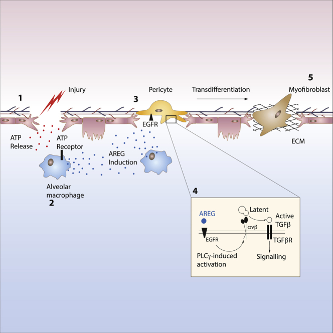
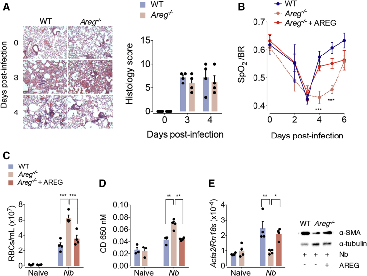
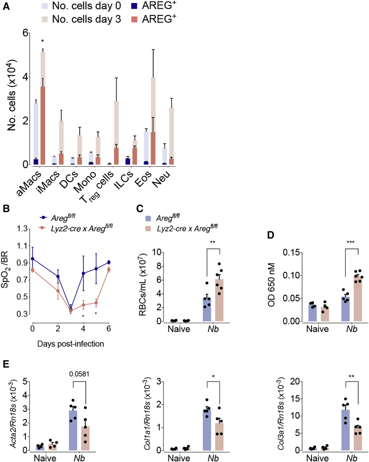
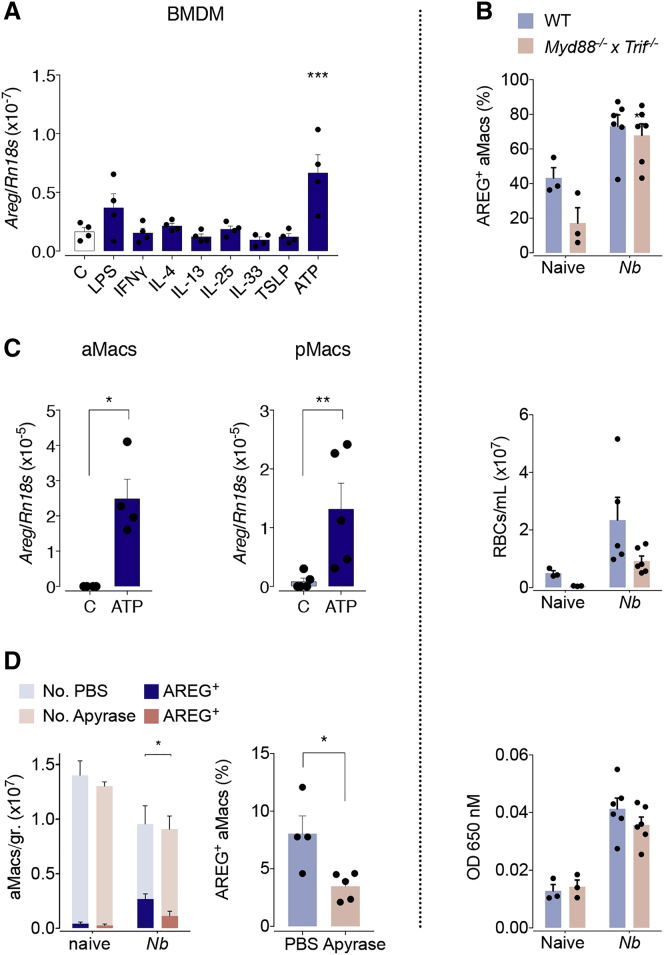
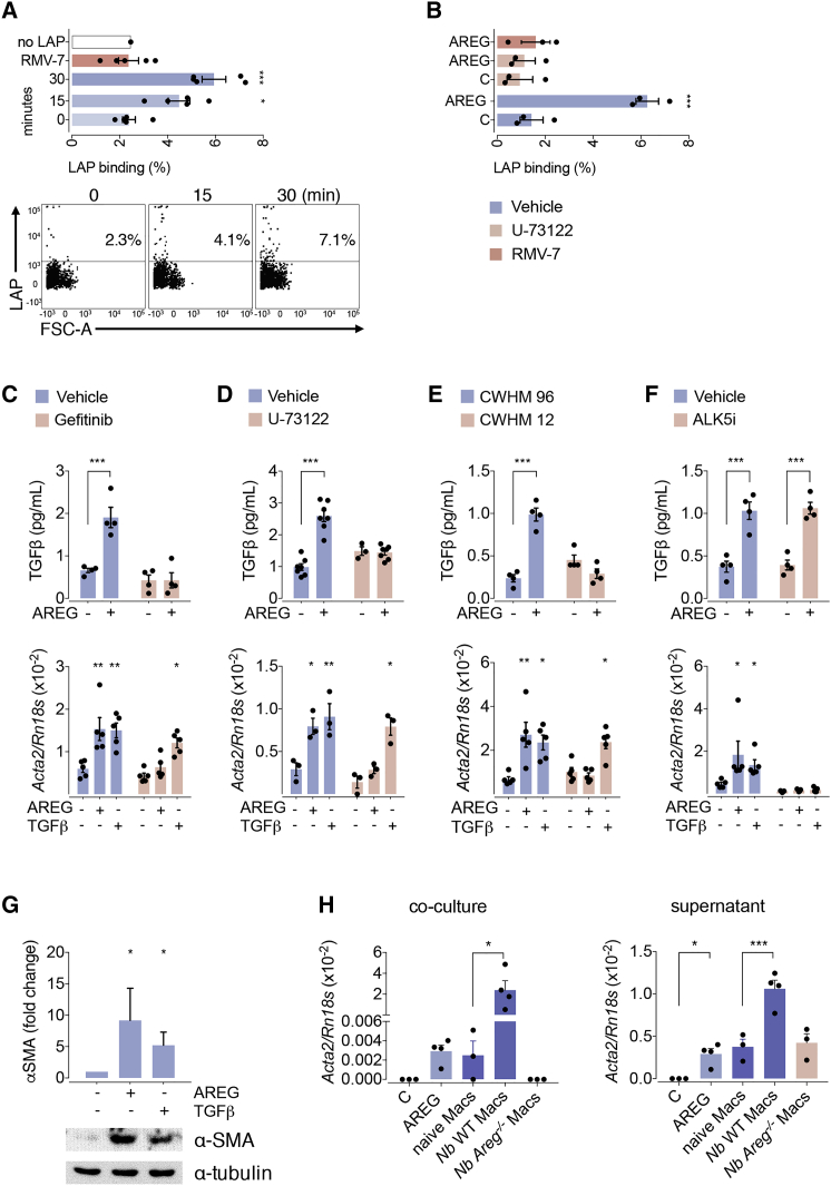
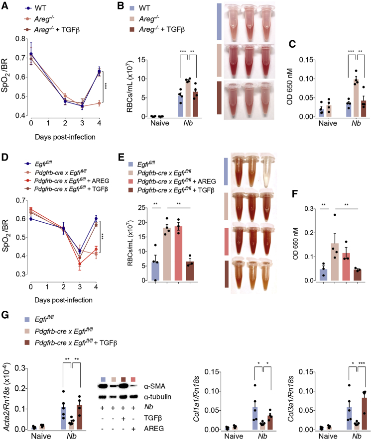
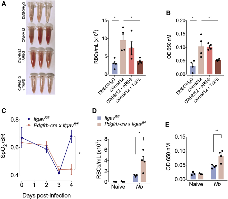
The epidermal growth factor receptor ligand Amphiregulin has a well-documented role in the restoration of tissue homeostasis after injury; however, the mechanism by which Amphiregulin contributes to wound repair remains unknown. Here we show that Amphiregulin functioned by releasing bioactive transforming growth factor beta (TGF-β) from latent complexes via integrin-αV activation. Using acute injury models in two different tissues, we found that by inducing TGF-β activation on mesenchymal stromal cells (pericytes), Amphiregulin induced their differentiation into myofibroblasts, thereby selectively contributing to the restoration of vascular barrier function within injured tissue. Furthermore, we identified macrophages as a critical source of Amphiregulin, revealing a direct effector mechanism by which these cells contribute to tissue restoration after acute injury. Combined, these observations expose a so far under-appreciated mechanism of how cells of the immune system selectively control the differentiation of tissue progenitor cells during tissue repair and inflammation.

Keywords: Amphiregulin; Macrophages; TGFb; pericytes.


Figures

None
Graphical abstract
Figure 1
Figure 1
Amphiregulin Contributes to the Restoration of Blood Barrier and Lung Function WT and Areg−/− mice were either left uninfected or infected with N. brasiliensis and either injected with 5 μg of rAREG at days 1, 2, and 3 post infection or left untreated. (A) Representative H&E staining and histological analysis of lung tissue at different dpi (days post infection). (B) Oxygen saturation in the blood at different dpi. (C) Number of red blood cells in the BAL (bronchoalveolar lavage). (D) Extravasation of Evans blue into the alveolar space as a marker of vascular permeability. (E) mRNA (Acta2) and protein expression of the αSMA at 4 dpi were evaluated by qRT-PCR and WB. All data are representative of at least two independent experiments (mean ± SEM); results for individual mice are shown as dots. See also Figure S1.
Figure 2
Figure 2
Macrophage-Derived Amphiregulin Contributes to the Restoration of Blood Barrier Function WT and Aregflox/flox × Lyz2cre mice were either left uninfected or infected with N. brasiliensis. (A) Absolute number of Amphiregulin expressing leukocytes following brefeldin A injection and intracellular cytokine staining of whole lung lysates at 3 dpi. (B) Oxygen saturation in the blood at different dpi. (C) Number of red blood cells in the BAL. (D) Extravasation of Evans blue into the alveolar space. (E) Expression of αSMA and collagen α1 type-I- and type-III-encoding genes at 4 dpi were evaluated. All data are representative of at least two independent experiments (mean ± SEM); results for individual mice are shown as dots. See also Figure S2.
Figure 3
Figure 3
Sensing Extracellular ATP from Tissue Necrosis Drives Amphiregulin Expression by Macrophages (A) In vitro differentiated bone marrow derived macrophages were treated as indicated. Expression of Amphiregulin-encoding gene was measured 10 h after treatment. (B) WT and Myd88−/− × Trif−/− mice were either left uninfected or infected with N. brasiliensis. Percentage of Amphiregulin-expressing (cell surface and intracellular after i.v. injection of brefeldin A) macrophages at 3 dpi (upper panel), number of red blood cells in the BAL (middle panel), and extravasation of Evans blue into the alveolar space at 4 dpi (lower panel). (C) Alveolar and peritoneal macrophages were purified by adherence and then treated with ATP. Expression of Amphiregulin-encoding gene was measured 10 h after treatment. (D) WT mice were either left uninfected or infected with N. brasiliensis and either received two individual doses of Apyrase at day 1 post-infection, or did not. Number and percentage of Amphiregulin expressing alveolar macrophages following brefeldin A injection and intracellular cytokine staining of lung lysate at 2 dpi. All data are representative of at least two independent experiments (mean ± SEM); results for individual mice are shown as dots. See also Figure S3.
Figure 4
Figure 4
Amphiregulin Induces Pericyte Differentiation by Releasing Bioactive TGF-β via Integrin-αV (A and B) Primary lung pericytes were cultured in the presence or absence of 100 ng/ml Amphiregulin (A), then incubated with latent TGF-β in the presence and absence of PLCγ inhibitor U-73122 (B) and analyzed for LAP binding by flow cytometry. (C–F) After 24 h of treatment, the release of bioactive TGF-β (upper panel), as well as their differentiation into myofibroblasts (lower panels), was determined in the presence or absence of inhibitors for the EGFR (Gefitinib) (C), PLCγ inhibitor (U-73122) (D), integrin-αV (CWHM-12 and its inactive control enantiomer CWHM-96) (E), or TGF-β-R (ALK5i) (F). (G) The induction of αSMA in treated pericytes was also evaluated at the protein level by western blot analysis. (H) Differentiation of primary lung pericytes into myofibroblasts following o/n co-culture with (left graph) or o/n exposure to supernatants derived from (right graph) alveolar macrophages isolated from infected or uninfected WT or Areg-/- mice. All data are representative of at least two independent experiments except for (B)–(D) (mean ± SEM); results for preparations from individual mice are shown as dots. See also Figure S4.
Figure 5
Figure 5
r TGF-β Restores Tissue Repair in Areg−/− Mice (A–C) WT or Areg−/− mice were either left uninfected or infected with N. brasiliensis. On days 1, 2, and 3 pi, mice were treated with 5 μg of rTGFβ or left untreated. (A) Oxygen saturation in the blood at different dpi, (B) number of red blood cells in the BAL, and (C) extravasation of Evans blue into the alveolar space at 4 dpi were evaluated. (D–G) Egfrflox/flox or Egfrflox/flox × Pdgfrb-cre mice were either left uninfected or infected with N. brasiliensis. On days 1, 2, and 3 pi, mice were treated with 5 μg of either rAREG or rTGFβ or left untreated. (D) Oxygen saturation in the blood at different dpi, (E) number of red blood cells in the BAL, (F) extravasation of Evans blue into the alveolar space, and (G) expression of the αSMA and collagen α1 types I and III were evaluated at 4 dpi. Data represent mean ± SEM; results for individual mice are shown as dots. See also Figure S5.
Figure 6
Figure 6
Amphiregulin Restores Blood Barrier Function via Pericyte-Specific Activation of Integrin-αV Complexes (A and B) WT mice were infected with N. brasiliensis or left uninfected, and minipumps containing the integrin-αV inhibitor CWHM12 were inserted subcutaneously 3 days prior to infection. Mice were treated with 5 μg of either rAREG or rTGFβ 1, 2, and 3 dpi. (A) Number of red blood cells in the BAL and (B) extravasation of Evans blue into the alveolar space were evaluated 4 dpi. (C–E) Igtavflox/flox and Igtavflox/flox × Pdgfrb-cre mice were infected with N. brasiliensis or left uninfected. (C) Oxygen saturation in the blood at different dpi. (D) Number of red blood cells in the BAL and (E) extravasation of Evans blue into the alveolar space were evaluated 4 dpi. Data represent mean ± SEM; results for individual mice are shown as dots. See also Figure S6.

References

    1. Abe M., Harpel J.G., Metz C.N., Nunes I., Loskutoff D.J., Rifkin D.B. An assay for transforming growth factor-beta using cells transfected with a plasminogen activator inhibitor-1 promoter-luciferase construct. Anal. Biochem. 1994;216:276–284. - PubMed
    1. Adachi O., Kawai T., Takeda K., Matsumoto M., Tsutsui H., Sakagami M., Nakanishi K., Akira S. Targeted disruption of the MyD88 gene results in loss of IL-1- and IL-18-mediated function. Immunity. 1998;9:143–150. - PubMed
    1. Arpaia N., Green J.A., Moltedo B., Arvey A., Hemmers S., Yuan S., Treuting P.M., Rudensky A.Y. A Distinct Function of Regulatory T Cells in Tissue Protection. Cell. 2015;162:1078–1089. - PMC - PubMed
    1. Aurora A.B., Olson E.N. Immune modulation of stem cells and regeneration. Cell Stem Cell. 2014;15:14–25. - PMC - PubMed
    1. Basak O., Beumer J., Wiebrands K., Seno H., van Oudenaarden A., Clevers H. Induced Quiescence of Lgr5+ Stem Cells in Intestinal Organoids Enables Differentiation of Hormone-Producing Enteroendocrine Cells. Cell Stem Cell. 2017;20:177–190.e4. - PubMed

Publication types

Substances