Review
. 2019 May;25(5):539-556.
doi: 10.1261/rna.070169.118. Epub 2019 Feb 15.

The search for a PKR code-differential regulation of protein kinase R activity by diverse RNA and protein regulators

Affiliations
Review

The search for a PKR code-differential regulation of protein kinase R activity by diverse RNA and protein regulators

Charles Bou-Nader et al. RNA. 2019 May.

Abstract

The interferon-inducible protein kinase R (PKR) is a key component of host innate immunity that restricts viral replication and propagation. As one of the four eIF2α kinases that sense diverse stresses and direct the integrated stress response (ISR) crucial for cell survival and proliferation, PKR's versatile roles extend well beyond antiviral defense. Targeted by numerous host and viral regulators made of RNA and proteins, PKR is subject to multiple layers of endogenous control and external manipulation, driving its rapid evolution. These versatile regulators include not only the canonical double-stranded RNA (dsRNA) that activates the kinase activity of PKR, but also highly structured viral, host, and artificial RNAs that exert a full spectrum of effects. In this review, we discuss our deepening understanding of the allosteric mechanism that connects the regulatory and effector domains of PKR, with an emphasis on diverse structured RNA regulators in comparison to their protein counterparts. Through this analysis, we conclude that much of the mechanistic details that underlie this RNA-regulated kinase await structural and functional elucidation, upon which we can then describe a "PKR code," a set of structural and chemical features of RNA that are both descriptive and predictive for their effects on PKR.

Keywords: PKR; allosteric regulation; antiviral defense; innate immunity; noncoding RNA.


Figures

FIGURE 1.
FIGURE 1.
Structural organization of PKR and schematic representation of different activation mechanisms. (A) Different domains of PKR: dsRBM1 is shown in blue, dsRBM2 in red, the basic patch at the amino terminus of the KD in purple, and the Ser/Thr KD in green with a smaller N-lobe than C-lobe. (B) Superposition of the different NMR conformers of PKR dsRBMs (pdb 1qu6) on dsRBM1 as reference (left) or on dsRBM2 (right), showing the dynamic nature of their intervening linker. Colored as in panel A. (C) Activate dimeric KD structure of PKR as seen in complex with eIF2α (pdb 2a1a and 2a19). The nonhydrolyzable ATP analog (AMP-PNP) is represented in sticks and the phosphorylated T446 in spheres. The N-lobe is in wheat and the C-lobe in green. Helix αC (involved in allosteric regulation) is highlighted in magenta and helix αG (involved in eIF2α recognition) is in red, while the activation loop is in dark blue and the P + 1 loop in cyan. (D) Dimeric KD of PKR K296R mutant (pdb 3uiu) colored as in C. Wild-type and K296R PKR share a similar dimeric interface, with the following exception: P-T446, absent from K296R, is required for stabilization/folding of the P + 1 loop for proper substrate recognition. This involves a network of interaction ranging from helix αC recognizing P-T446 further rigidifying the activation loop, allowing for P + 1 loop folding and its interaction with helix αG. (E) PKR is proposed to exist in a multitude of states with distinct structures and activities. PKR phosphorylation following activation is indicated with a yellow star (phosphorylation of T446 or dsRBM1). In its inactive state, PKR is mainly monomeric either extended in solution or locked in an auto-inhibited state with dsRBM2 bound to the KD. Two different dsRBM2-binding interfaces on KD have been proposed; one involving the N-lobe while the other was mapped at the C-lobe next to helix αG. Binding to >30 bp dsRNA induces PKR activation either by relieving the auto-inhibited state and/or by dimerization of PKR on the same RNA, bringing two KDs in close proximity. PACT is thought to bind PKR, especially via its dsRBM3 (M3, see below), and activates KD through a similar mechanism. Shorter dsRNA length induces PKR dimerization but no activation. This could imply that dsRBM2 remains bound to the KD while only dsRBM1 interacts with the RNA and/or assists in forming inactive dimers. Following activation of PKR by different stimuli, T446 is phosphorylated and both dimeric and monomeric activated PKR have been observed, leading to efficient eIF2α phosphorylation. Subsequently, phosphorylation of dsRBM1 could gradually inactivate PKR by interacting with the KD returning to an auto-inhibited state post-activation.
FIGURE 2.
FIGURE 2.
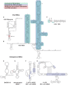
Secondary structures of several naturally occurring RNA activators of PKR. (A) HDV ribozyme, (B) HCV IRES, (C) HIV-1 Tar, (D) SNORD113, (E) human mt-tRNALeu, (F) 3′-UTR 2-APRE of TNF-α pre-Mrna, and (G) 5′-UTR IFN-γ pre-mRNA. Regions relevant to PKR binding are highlighted in cyan, where mutations lead to decreased affinity of PKR. Regions relevant to PKR activation are highlighted in blue, where mutations lead to decreased activation of PKR. Regions that enhance PKR activation by inducing RNA dimerization are highlighted in red.
FIGURE 3.
FIGURE 3.
Secondary structures of PKR-inhibitory RNAs. (A) VA-I, (B) EBER-1, and (C) nc886. Regions involved in PKR binding are highlighted in cyan while those required for inhibition are in blue.
FIGURE 4.
FIGURE 4.
Schematic representation of protein regulators of PKR with their interacting interfaces. Blue lines represent interactions with PKR dsRBMs, while gray lines denote interactions with the Ser/Thr KD. Dashed lines highlight direct competition for dsRNA binding. The domain organization of PKR protein regulators and their boundaries are represented following the color code: Class A's in light blue, Class B's in red, Z-DNA binding domain in yellow, DZF dimerization domain in purple, A to I deaminase catalytic domain in light green, dihydrouridine synthase conserved catalytic domain in orange, Zinc binding domain in pink, DnaJ domain in dark green, ISDR motif in dark gray, and TPR repeat motifs in light gray.

References

    1. Andersen ES, Contera SA, Knudsen B, Damgaard CK, Besenbacher F, Kjems J. 2004. Role of the trans-activation response element in dimerization of HIV-1 RNA. J Biol Chem 279: 22243–22249. 10.1074/jbc.M314326200 - DOI - PubMed
    1. Anderson E, Cole JL. 2008. Domain stabilities in protein kinase R (PKR): evidence for weak interdomain interactions. Biochemistry 47: 4887–4897. 10.1021/bi702211j - DOI - PMC - PubMed
    1. Ben-Asouli Y, Banai Y, Pel-Or Y, Shir A, Kaempfer R. 2002. Human interferon-γ mRNA autoregulates its translation through a pseudoknot that activates the interferon-inducible protein kinase PKR. Cell 108: 221–232. 10.1016/S0092-8674(02)00616-5 - DOI - PubMed
    1. Benkirane M, Neuveut C, Chun RF, Smith SM, Samuel CE, Gatignol A, Jeang KT. 1997. Oncogenic potential of TAR RNA binding protein TRBP and its regulatory interaction with RNA-dependent protein kinase PKR. EMBO J 16: 611–624. 10.1093/emboj/16.3.611 - DOI - PMC - PubMed
    1. Bevilacqua PC, George CX, Samuel CE, Cech TR. 1998. Binding of the protein kinase PKR to RNAs with secondary structure defects: role of the tandem A-G mismatch and noncontiguous helixes. Biochemistry 37: 6303–6316. 10.1021/bi980113j - DOI - PubMed

Publication types

MeSH terms