. 2019 Apr 4;85(8):e02133-18.
doi: 10.1128/AEM.02133-18. Print 2019 Apr 15.

The Edwardsiella piscicida Type III Translocon Protein EseC Inhibits Biofilm Formation by Sequestering EseE

Affiliations

The Edwardsiella piscicida Type III Translocon Protein EseC Inhibits Biofilm Formation by Sequestering EseE

Ying Li Liu et al. Appl Environ Microbiol. .

Abstract

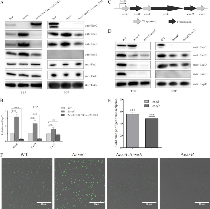
The type III secretion system (T3SS) is one of the most important virulence factors of the fish pathogen Edwardsiella piscicida It contains three translocon proteins, EseB, EseC, and EseD, required for translocation of effector proteins into host cells. We have previously shown that EseB forms filamentous appendages on the surface of E. piscicida, and these filamentous structures mediate bacterial cell-cell interactions promoting autoaggregation and biofilm formation. In the present study, we show that EseC, but not EseD, inhibits the autoaggregation and biofilm formation of E. piscicida At 18 h postsubculture, a ΔeseC strain developed strong autoaggregation and mature biofilm formation, accompanied by enhanced formation of EseB filamentous appendages. This is in contrast to the weak autoaggregation and immature biofilm formation seen in the E. piscicida wild-type strain. EseE, a protein that directly binds to EseC and also positively regulates the transcription of the escC-eseE operon, was liberated and showed increased levels in the absence of EseC. This led to augmented transcription of the escC-eseE operon, thereby increasing the steady-state protein levels of intracellular EseB, EseD, and EseE, as well as biofilm formation. Notably, the levels of intracellular EseB and EseD produced by the ΔeseE and ΔeseC ΔeseE strains were similar but remarkably lower than those produced by the wild-type strain at 18 h postsubculture. Taken together, we have shown that the translocon protein EseC inhibits biofilm formation through sequestering EseE, a positive regulator of the escC-eseE operon.IMPORTANCEEdwardsiella piscicida, previously known as Edwardsiella tarda, is a Gram-negative intracellular pathogen that mainly infects fish. The type III secretion system (T3SS) plays a pivotal role in its pathogenesis. The T3SS translocon protein EseB is required for the assembly of filamentous appendages on the surface of E. piscicida The interactions between the appendages facilitate autoaggregation and biofilm formation. In this study, we explored the role of the other two translocon proteins, EseC and EseD, in biofilm formation. We have demonstrated that EseC, but not EseD, inhibits the autoaggregation and biofilm formation of E. piscicida, providing new insights into the regulatory mechanism involved in E. piscicida biofilm formation.

Keywords: Edwardsiella piscicida; biofilm; translocon protein; type III secretion system.


Figures

FIG 1
FIG 1
Dynamic analysis of E. piscicida autoaggregation. E. piscicida strains were cultured in DMEM in glass tubes. Autoaggregation was recorded at 18 h and 24 h postsubculture (hps).
FIG 2
FIG 2
T3SS translocon protein EseC inhibits the biofilm formation of E. piscicida at 18 h postsubculture. (A) E. piscicida strains were subcultured in DMEM in a 24-well plate embedded with coverslips. At 18 h and 24 h postsubculture, bacteria settled onto the coverslips were fixed and stained with 0.1% crystal violet. Quantification of dissolved crystal violet was performed to evaluate the biofilm formation ability. ***, P < 0.001; NS, not significant. (B) Immunofluorescence staining on WT and ΔeseC mutant strains with an antibody against EseB. E. piscicida WT and ΔeseC mutant strains were subcultured in DMEM in a 24-well plate embedded with coverslips. Images were taken using a confocal laser scanning microscope. Scale bar = 5 μm.
FIG 3
FIG 3
Intracellular EseB production is decreased in the presence of EseC. (A) The TBP from equal amounts of E. piscicida wild-type, ΔeseC, and ΔeseC/pJN-eseC strains were probed with antibodies against EseC, EseB, and EvpC. EvpC, a T6SS-secreted protein, was used as the loading control. To induce the expression of EseC, 10 mM l-arabinose was supplemented into the culture of the ΔeseC/pJN-eseC complementation strain. (B) Quantitative analysis of EseB protein level from TBP. Proteins were quantified by densitometry and normalized to EvpC. The graphs show the relative ratios of intracellular EseB, which are averages of the results from at least three independent experiments. **, P < 0.01.
FIG 4
FIG 4
The loss of EseC frees up EseE that upregulates the transcription of escC-eseE operon. (A) The TBP and ECP from equal amounts of E. piscicida strains collected at 18 h postsubculture were probed with antibodies against EseC, EseB, EseD, EseE, EseG, EseJ, and EvpC. EvpC, a T6SS-secreted protein, was used as the loading control. The immunoblotting data shown are representative of three independent experiments. (B) Quantitative analysis of protein levels of EseB, EseD, and EseE from bacterial pellets. Proteins were quantified by densitometry and normalized to EvpC. The graphs show the relative ratios of intracellular EseB, EseD, and EseE, which are averages of the results from at least three independent experiments. ***, P < 0.001; **, P < 0.01; NS, not significant. (C) Schematic description of the escC to eseG region of the E. piscicida T3SS. Each arrow represents an open reading frame. The bent arrow represents the putative promoter region. (D) The loss of EseE nullifies the inhibitory effect of EseC on the production of (intracellular and extracellular) EseB. At 18 h postsubculture, TBP and ECP from equal amounts of different E. piscicida strains were probed with antibodies against EseC, EseB, EseD, EseE, and EvpC. EvpC was also used as the loading control. The immunoblotting data shown are representative of three independent experiments. (E) EseE regulates the transcription of eseB and eseD. The mRNA levels of eseB and eseD in the ΔeseC and ΔeseC ΔeseE mutant strains were examined by quantitative PCR (qPCR). Gene expression levels in the ΔeseC strain relative to those in the ΔeseC ΔeseE strain are presented. 16S rRNA was used as the reference gene. Data are presented as the mean ± SEM from three independent experiments. ***, P < 0.001. (F) A DNA fragment (bp −200 to −1) upstream of escC was inserted into the promoterless gfp shuttle vector pFPV25 to create pFPV−200–−1. This construct (pFPV−200–−1) was introduced into E. piscicida WT and ΔeseC, ΔeseC ΔeseE, ΔesrB, and ΔesrC strains. At 18 h postsubculture in DMEM, the images of those E. piscicida strains were taken using a confocal microscope. Scale bar = 50 μm.
FIG 5
FIG 5
EseC inhibits biofilm formation through sequestering EseE. E. piscicida strains were subcultured in DMEM in a 24-well plate embedded with coverslips. At 18 h and 24 h postsubculture, bacteria settled onto the coverslips were fixed and stained with 0.1% crystal violet. Quantification of dissolved crystal violet was performed to evaluate the biofilm formation ability of the bacteria. ***, P < 0.001; *, P < 0.05; NS, not significant.

References

    1. Ling SH, Wang XH, Xie L, Lim TM, Leung KY. 2000. Use of green fluorescent protein (GFP) to study the invasion pathways of Edwardsiella tarda in in vivo and in vitro fish models. Microbiology 146:7–19. doi:10.1099/00221287-146-1-7. - DOI - PubMed
    1. Shao S, Lai QL, Liu Q, Wu HZ, Xiao JF, Shao ZZ, Wang QY, Zhang YX. 2015. Phylogenomics characterization of a highly virulent Edwardsiella strain ET080813T encoding two distinct T3SS and three T6SS gene clusters: propose a novel species as Edwardsiella anguillarum sp. nov. Syst Appl Microbiol 38:36–47. doi:10.1016/j.syapm.2014.10.008. - DOI - PubMed
    1. Miwa S, Mano N. 2000. Infection with Edwardsiella tarda causes hyperthrophy of liver cells in the Japanese flounder Paralichthys olivaceus. Dis Aquat Org 42:227–231. doi:10.3354/dao042227. - DOI - PubMed
    1. Padrós F, Zarza C, Dopazo L, Cuadrado M, Crespo S. 2006. Pathology of Edwardsiella tarda infection in turbot, Scophthalmus maximus (L.). J Fish Dis 29:87–94. doi:10.1111/j.1365-2761.2006.00685.x. - DOI - PubMed
    1. Tan YP, Zheng J, Tung SL, Rosenshine I, Leung KY. 2005. Role of type III secretion in Edwardsiella tarda virulence. Microbiology 151:2301–2313. doi:10.1099/mic.0.28005-0. - DOI - PubMed

Publication types

MeSH terms

Supplementary concepts

LinkOut - more resources