. 2019 Feb 15;10(3):162.
doi: 10.1038/s41419-019-1388-5.

NETO2 promotes invasion and metastasis of gastric cancer cells via activation of PI3K/Akt/NF-κB/Snail axis and predicts outcome of the patients

Affiliations

NETO2 promotes invasion and metastasis of gastric cancer cells via activation of PI3K/Akt/NF-κB/Snail axis and predicts outcome of the patients

Jun-Yan Liu et al. Cell Death Dis. .

Abstract

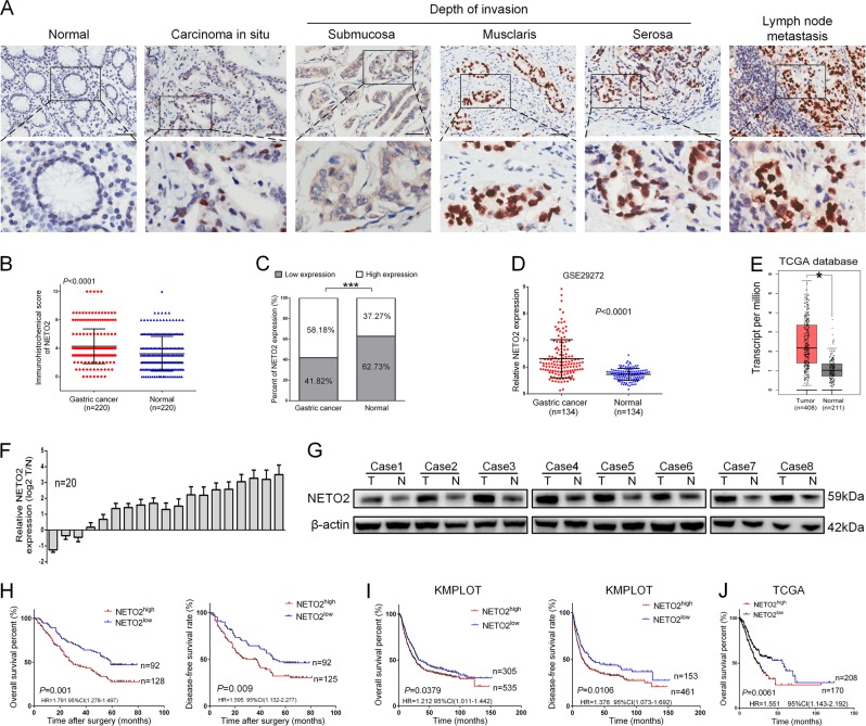
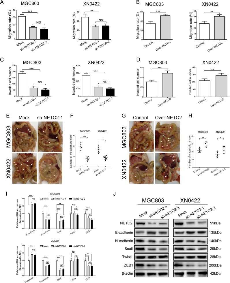
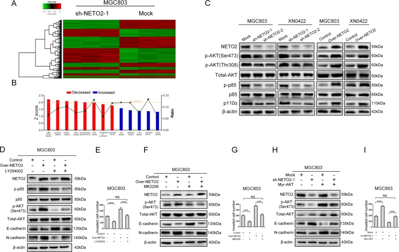
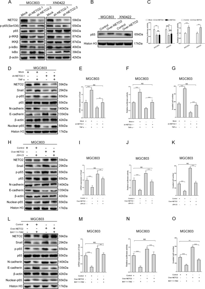
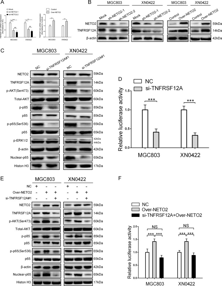
Aberrant expression of neuropilin and tolloid-like 2 (NETO2) has been observed during the progression of some human carcinomas. However, the expression pattern and clinical relevance of NETO2 in gastric cancer (GC) remain to be elucidated. In this study, we found that NETO2 expression was higher in GC tissues compared with paired non-cancerous tissues. Moreover, the expression of NETO2 was positively correlated with clinical stage, invasion depth, lymph node metastasis, and tumor size, but inversely correlated with overall and disease-free survival rates. Cox regression analysis identified NETO2 as an independent prognostic indicator for GC patients. Overexpression of NETO2 facilitated migration and invasion of GC cells in vitro and metastasis in vivo in association with induction of epithelial-mesenchymal transition. Conversely, knockdown of NETO2 had the opposite effects. Mechanistically, silencing NETO2 reduced the phosphorylation of PI3K, AKT, and NF-κB p65 as well as the expression of Snail, whereas NETO2 overexpression achieved the opposite results. Furthermore, we identified TNFRSF12A as a mediator for NETO2 to activate PI3K/AKT/NF-κB/Snail axis. Collectively, our results demonstrate that NETO2 promotes invasion and metastasis of GC cells and represents a novel prognostic indicator as well as a potential therapeutic target in GC.


Conflict of interest statement

The authors declare that they have no conflict of interest.

Figures

Fig. 1
Fig. 1. NETO2 is highly expressed in gastric cancer tissues and correlated with outcome of the patients.
a Representative immunohistochemical staining (IHC) images of NETO2 in adjacent normal tissue, gastric cancer tissues with different invasion depth and metastatic lymph node. Scale bar = 50 μm. b The IHC score of NETO2 in gastric cancer tissues was significantly higher than that in adjacent normal tissues. c High expression of NETO2 was more frequent in gastric cancer tissues than in adjacent normal tissues. d Data in GEO GSE29272 showed higher mRNA level of NETO2 in gastric cancer tissues compared to adjacent normal tissues. e Data in TCGA database showed higher mRNA level of NETO2 in gastric cancer tissues compared to adjacent normal tissues. f mRNA levels of NETO2 were higher in 20 fresh surgical gastric tumor specimens (t) than that in paired adjacent normal tissues (n). g NETO2 protein levels were higher in eight fresh surgical gastric cancer (t) tissues than that in adjacent normal (n) tissues. h Kaplan–Meier estimation indicated significantly lower overall and disease-free survival rates in patients with NETO2high gastric cancer than that in NETO2low patients. i Data in KMPLOT database showed that NETO2high patients had significantly lower overall and disease-free survival rates than that in NETO2low patients. j Data in TCGA database showed that NETO2high patients had significantly lower overall survival rate than that in NETO2low patients. *p < 0.05, ***p < 0.001
Fig. 2
Fig. 2. NETO2 promotes the migration and invasion of gastric cancer cells in vitro and metastasis in vivo in association with epithelial–mesenchymal transition (EMT).
a Quantification of wound healing assay showed decreased migration ability in NETO2-knockdown cells compared to mock cells. b Quantification of wound healing assay showed increased migration ability in Over-NETO2 cells compared to control cells. c Quantification of transwell invasion assay showed decreased invasive ability in NETO2-knockdown cells compared to mock cells. d Quantification of transwell invasion assay showed increased invasive ability in Over-NETO2 cells compared to control cells. e Representative images of intraperitoneal metastasis assay showed metastatic nodules derived from sh-NETO2–1 cells and mock cells. Arrows indicate intraperitoreal nodules. f Quantification of intraperitoneal metastasis assay showed reduced number of metastatic foci formed by NETO2-knockdown cells compared to mock cells. g Representative images of intraperitoneal metastasis assay showed metastatic nodules derived from Over-NETO2 and control cells. Arrows indicate intraperitoreal nodules. h Quantification of intraperitoneal metastasis assay showed increased number of metastatic foci formed by Over-NETO2 cells compared to control cells. i qRT-PCR analysis showed significantly upregulated E-cadherin and downregulated N-cadherin, Snail and ZEB1 at mRNA level in NETO2-knockdown cells compared to mock cells. j Western blotting analysis showed significantly upregulated E-cadherin and downregulated Fibronectin, N-cadherin, Snail, and ZEB1 at protein level in NETO2-knockdown cells compared to mock cells. *p < 0.05, **p < 0.01, ***p < 0.001, NS no significant
Fig. 3
Fig. 3. NETO2 activates PI3K/AKT pathway to induce EMT in GC cells.
a Microarray heatmap showed differentially expressed genes in mock and sh-NETO2–1 cells. The color key for the normalized expression data was shown at the top of the microarray heatmap (green means downregulated genes and red represents upregulated genes). b Ingenuity Pathway Analysis (IPA) of the most significantly changed classical pathways after knockdown of NETO2 in gastric cancer cells. All signaling pathways were sorted according to the z score (left y-axis). The ratio represented the relative number of differentially expressed genes in a specified signaling pathway (right y-axis). c Western blotting analysis showed reduced phosphorylation levels of AKT and p85 in sh-NETO2–1 cells, while increased phosphorylation levels of AKT and p85 in Over-NETO2 cells compared to their control cells, respectively. d Western blotting analysis showed that treatment with LY294002 (10 μM) attenuated NETO2-enhanced phosphorylation of AKT and inhibited NETO2-induced EMT in Over-NETO2 cells. e Quantification of transwell invasion assay showed that treatment with LY294002 (10 μM) attenuated the enhanced invasive ability in Over-NETO2 cells. f Western blotting analysis showed that treatment with MK2206 (5 μM) attenuated NETO2-enhanced phosphorylation of AKT and inhibited NETO2-induced EMT in Over-NETO2 cells. g Quantification of transwell invasion assay showed that treatment with MK2206 (5 μM) attenuated the invasive ability in Over-NETO2 cells. h Western blotting analysis showed that transfection with myristoylated (myr) -AKT increased the phosphorylation level of AKT and induced EMT in NETO2-knockdown cells. i Quantification of transwell invasion assay showed that transfection with myr-AKT increased the invasive ability in NETO2-knockdown cells. ***p<0.001; NS no significant
Fig. 4
Fig. 4. PI3K/AKT activates NF-κB/Snail axis to regulate NETO2-induced EMT.
a Western blotting analysis showed attenuated phosphorylation of p65, IKKβ, and IκBα in NETO2-knockdown cells compared to mock cells. b Western blotting analysis showed increased nuclear accumulation of NF-κB p65 in Over-NETO2 cells compared to control cells. c The NF-κB transcription activity was decreased in NETO2-knockdown gastric cancer cells, while increased in NETO2-overexpression gastric cancer cells. d Western blotting analysis showed that TNF-α (10 ng/mL) treatment resulted in increased phosphorylation and nuclear accumulation of p65, downregulated E-cadherin and upregulated N-cadherin in sh-NETO2–1 cells. e The mRNA level of Snail was upregulated after treatment with TNF-α (10 ng/mL) in sh-NETO2–1 GC cells. f Treatment with TNF-α (10 ng/mL) enhanced NF-κB transcription activity measured by dual luciferase assay in sh-NETO2–1 GC cells. g Quantification of transwell invasion assay showed increased invasive ability after treatment with TNF-α (10 ng/mL) in sh-NETO2–1 GC cells. h Western blotting analysis showed that JSH-23 (10 μM) treatment led to decreased phosphorylation and nuclear accumulation of p65, upregulated E-cadherin, and downregulated N-cadherin in Over-NETO2 cells. i The mRNA level of Snail was decreased after treatment with JSH-23 (10 μM) in Over-NETO2 GC cells. j Treatment with JSH-23 (10 μM) decreased the NF-κB transcription activity measured by dual luciferase assay in Over-NETO2 GC cells. k Quantification of transwell invasion assay showed decreased invasive ability after treatment with JSH-23 (10 μM) in Over-NETO2 GC cells. l Western blotting analysis showed that BAY 11–7082 (10 μM) treatment resulted in decreased phosphorylation and nuclear accumulation of p65, upregulated E-cadherin and downregulated N-cadherin in Over-NETO2 cells. m The mRNA level of Snail was decreased after treatment with BAY 11–7082 (10 μM) in Over-NETO2 GC cells. n Treatment with BAY 11–7082 (10 μM) decreased the NF-κB transcription activity measured by dual luciferase assay in Over-NETO2 GC cells. o Quantification of transwell invasion assay showed decreased invasive ability after treatment with BAY 11–7082 (10 μM) in Over-NETO2 GC cells. **p < 0.01, ***p < 0.001; NS no significant
Fig. 5
Fig. 5. TNFRSF12A mediates NETO2-induced activation of PI3K/AKT/NF-κB/Snail axis.
a qRT-PCR analysis showed knockdown of NETO2 downregulated the mRNA level of TNFRSF12A, while overexpression of NETO2 upregulated the mRNA level of TNFRSF12A. b Western blotting analysis showed that depletion of NETO2 downregulated TNFRSF12A expression, while overexpression of NETO2 upregulated TNFRSF12A expression. c Depletion of TNFRSF12A in MGC803 and XN0422 cells inactivated PI3K/AKT/NF-κB pathway, but had no influence on Erk1/2 pathway. d Depletion of TNFRSF12A impaired the NF-κB transcription activity in MGC803 and XN0422 cells. e Silencing TNFRSF12A abrogated NETO2-induced activation of PI3K/AKT/NF-κB pathway. f Depletion of TNFRSF12A inhibited NETO2-induced NF-κB transcription activity; *p < 0.05, **p < 0.01, ***p < 0.001; NS no significant

References

    1. Siegel RL, Miller KD, Jemal A. Cancer statistics, 2015. CA Cancer J. Clin. 2015;65:5. doi: 10.3322/caac.21254. - DOI - PubMed
    1. Siegel RL, Miller KD, Jemal A. Cancer statistics, 2016. CA Cancer J. Clin. 2016;66:7. doi: 10.3322/caac.21332. - DOI - PubMed
    1. Siegel RL, Miller KD, Jemal A. Cancer statistics, 2017. CA Cancer J. Clin. 2017;67:7. doi: 10.3322/caac.21387. - DOI - PubMed
    1. Van Cutsem E, Sagaert X, Topal B, Haustermans K, Prenen H. Gastric cancer. Lancet. 2016;388:2654. doi: 10.1016/S0140-6736(16)30354-3. - DOI - PubMed
    1. Stohr H, Berger C, Frohlich S, Weber BH. A novel gene encoding a putative transmembrane protein with two extracellular CUB domains and a low-density lipoprotein class A module: isolation of alternatively spliced isoforms in retina and brain. Gene. 2002;286:223. doi: 10.1016/S0378-1119(02)00438-9. - DOI - PubMed

Publication types

MeSH terms