. 2019 Feb 15;10(3):147.
doi: 10.1038/s41419-019-1414-7.

Fhit-Fdxr interaction in the mitochondria: modulation of reactive oxygen species generation and apoptosis in cancer cells

Affiliations

Fhit-Fdxr interaction in the mitochondria: modulation of reactive oxygen species generation and apoptosis in cancer cells

Teresa Druck et al. Cell Death Dis. .

Abstract

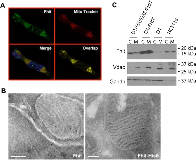
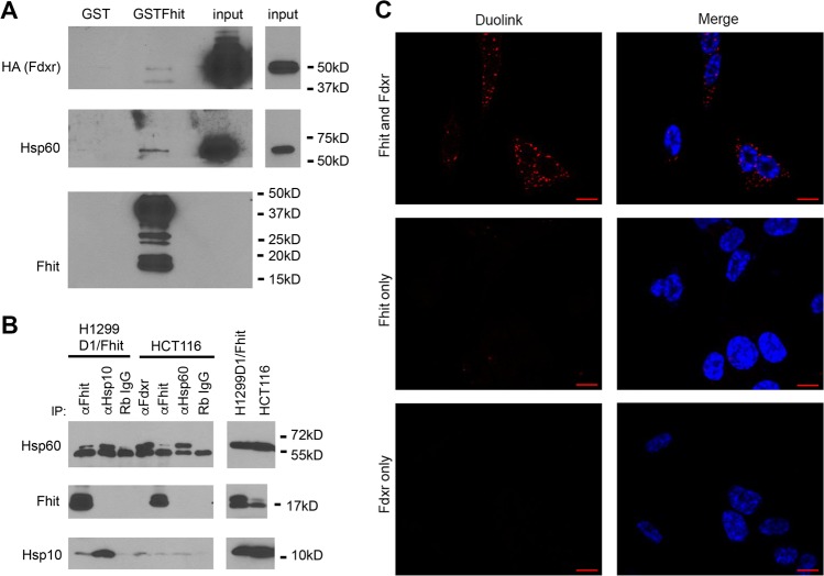
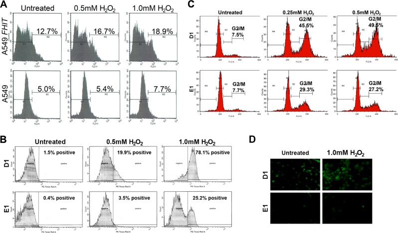
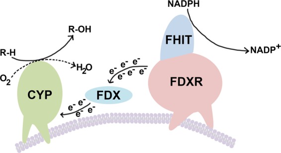
Fhit protein is lost in cancers of most, perhaps all, cancer types; when restored, it can induce apoptosis and suppress tumorigenicity, as shown in vitro and in mouse tumor models in vivo. Following protein cross-linking and proteomics analyses, we characterized a Fhit protein complex involved in triggering Fhit-mediated apoptosis. The complex includes the heat-shock chaperonin pair, HSP60/10, which is likely involved in importing Fhit into the mitochondria, where it interacts with ferredoxin reductase, responsible for transferring electrons from NADPH to cytochrome P450 via ferredoxin, in electron transport chain complex III. Overexpression of Fhit protein in Fhit-deficient cancer cells modulates the production of intracellular reactive oxygen species, causing increased ROS, following peroxide treatment, with subsequent increased apoptosis of lung cancer cells under oxidative stress conditions; conversely, Fhit-negative cells escape ROS overproduction and ROS-induced apoptosis, likely carrying oxidative damage. Thus, characterization of Fhit-interacting proteins has identified direct effectors of a Fhit-mediated apoptotic signal pathway that is lost in many cancers. This is of translational interest considering the very recent emphasis in a number of high-profile publications, concerning the role of oxidative phosphorylation in the treatment of human cancers, and especially cancer stem cells that rely upon oxidative phosphorylation for survival. Additionally, we have shown that cells from a Fhit-deficient lung cancer cell line, are sensitive to killing by exposure to atovaquone, thought to act as a selective oxidative phosphorylation inhibitor by targeting the CoQ10 dependence of the mitochondrial complex III, while the Fhit-expressing sister clone is resistant to this treatment.


Conflict of interest statement

The authors declare that they have no conflict of interest.

Figures

Fig. 1
Fig. 1. Subcellular localization of Fhit protein in the cytosol and mitochondria.
a Immunofluorescence microscopy was performed with anti-Fhit serum on H1299 cells (D1) treated with PonA for 8 h; Fhit staining was detected using fluorescein isothiocyanate- (green) conjugated anti-rabbit immunoglobulin (IgG); MitoTracker Red staining, which identifies mitochondria, shows partial colocalization with Fhit. The yellow color on the fourth panel (lower right) shows the co-localization points. b Immuno-electron microscopy of A549 AdFHIT (left) or AdFHIT-His6-infected cells (right) performed with a Penta-His antibody shows Fhit mitochondrial localization (right); A549 cells infected with AdFHIT served as a control and show only a few scattered grains (left). c Immunoblot analyses of cytosolic and mitochondrial protein fractions from H1299D1 and HCT116 cells. Vdac is a marker of the mitochondria
Fig. 2
Fig. 2. Fhit interacts with Fdxr, HSP10, and HSP60.
a GST pull-down experiment using a protein lysate from H1299D1 cells transfected with HA-FDXR which was incubated with GST and GST-Fhit protein bound to glutathione agarose resin, and protein complexes eluted and separated on acrylamide gel for detection with antisera against HA, HSP60, and Fhit. b Lysates were prepared from DSP-cross-linked H1299D1 cells (Pon A induced for Fhit expression) or from HCT116 cells (expressing endogenous Fhit) and used in IP experiments with the indicated antisera. Complexes were separated on acrylamide gel and proteins were detected with HSP60, Fhit, or HSP10 antisera. c The association of Fhit and Fdxr in HCT116 cells using the Duolink in situ proximity ligation assay (PLA). PLA signals are shown in red and nuclei in blue. PLA signals for Fhit/Fdxr are mostly confined in the cytoplasm. Negative controls with the Fdxr or Fhit antibody alone show no PLA signal. Scale bar represents 10 μm
Fig. 3
Fig. 3. Fhit expression induces intracellular ROS generation after treatment of cells with peroxide.
a FACS analysis for ROS assessment in A549 cells 48 h after transfection with FHIT plasmid, with or without 5 h of H2O2 treatment. Empty vector transfected cells served as a control. Intracellular superoxide was determined according to fluorescence of ethidium as a result of oxidation of hydroethidine by ·O2. M2 refers to the fraction of ROS-positive cells. b FACS analysis for ROS assessment by the fluorescence produced from the oxidation of hydroethidine in D1 and E1 cells; 48 h after PonA treatment, cells were treated for 5 h with 0.5 and 1.0 mM H2O2 and oxidative stress was measured; % positive refers to the fraction of fluorescent cells, indicating ROS. c FACS analysis of D1 and E1 cell cycle kinetics at 48 h after oxidative stress treatment. Cells were treated with PonA for 48 h and then with increasing concentrations of H2O2 (0.25, 0.5 mM) for 4 h. Analysis was at 48 h after H2O2 treatment. All experiments were performed twice in triplicate. d Increased green fluorescent DCF signal in H1299 Fhit-expressing cells (D1) under stress conditions. Cells were incubated with 2′,7′-dichlorodihydrofluorescein diacetate (H2DCFDA), a ROS indicator that can be oxidized in the presence of ROS to the highly green fluorescent dye dichlorofluorescein (DCF), at 48 h after Fhit induction and after 5 h of H2O2 treatment of E1 and D1 cells (×40 magnification)
Fig. 4
Fig. 4
Cell viability of Fhit-negative H1299 cells is decreased after atovaquone treatment. H1299 E1 and D1 cells were treated with atovaquone (10 μM) for 3 days. Cell viability was assessed by trypan blue staining. Data from three independent experiments. Note that the percentage of cell viability of atovaquone-exposed cells is normalized to the control DMSO-treated cells
Fig. 5
Fig. 5
Mitochondrial cytochrome P450 (cyp) ETC. The model is modified from Midzak & Papadopoulos. Membrane-bound ferredoxin reductase (Fdxr) accepts two electrons from NADPH, yielding NADP + ; these electrons are passed to the iron–sulfur cluster of ferredoxin (FDX), which donates the electrons to the heme prosthetic group of mitochondrial CYP, which uses protons and molecular O2 to hydroxylate its target substrate, yielding the final hydroxylated product, as described by Midzak & Papadopoulos. The electron donor, FDX, is a 14-kDa mitochondrial matrix protein containing a Fe–S cluster. The mechanism of Fhit action on the Fdx–Fdxr interaction is unknown but perhaps Fhit interrupts this interaction and thus blocks the passing of electrons to Fdx, allowing leakage of electrons and contribution to ROS production and apoptosis, under specific conditions

References

    1. Ohta M, et al. The FHIT gene, spanning the chromosome 3p14.2 fragile site and renal carcinoma-associated t(3;8) breakpoint, is abnormal in digestive tract cancers. Cell. 1996;84:587–597. doi: 10.1016/S0092-8674(00)81034-X. - DOI - PubMed
    1. Matsuyama A, et al. Fragile site orthologs FHIT/FRA3B and Fhit/Fra14A2: evolutionarily conserved but highly recombinogenic. Proc. Natl. Acad. Sci. U. S. A. 2003;100:14988–14993. doi: 10.1073/pnas.2336256100. - DOI - PMC - PubMed
    1. Karras JR, Schrock MS, Batar B, Huebner K. Fragile genes that are frequently altered in cancer: players not passengers. Cytogenet. Genome Res. 2016;150:208–216. doi: 10.1159/000455753. - DOI - PubMed
    1. Waters CE, Saldivar JC, Hosseini SA, Huebner K. The FHIT gene product: tumor suppressor and genome “caretaker”. Cell. Mol. Life Sci. 2014;71:4577–4587. doi: 10.1007/s00018-014-1722-0. - DOI - PMC - PubMed
    1. Volinia S, Druck T, Paisie CA, Schrock MS, Huebner K. The ubiquitous ‘cancer mutational signature’ 5 occurs specifically in cancers with deleted FHIT alleles. Oncotarget. 2017;8:102199–102211. doi: 10.18632/oncotarget.22321. - DOI - PMC - PubMed

Publication types

MeSH terms