. 2019 Sep;26(9):1545-1565.
doi: 10.1038/s41418-018-0270-9. Epub 2019 Feb 15.

Loss of glutathione redox homeostasis impairs proteostasis by inhibiting autophagy-dependent protein degradation

Affiliations

Loss of glutathione redox homeostasis impairs proteostasis by inhibiting autophagy-dependent protein degradation

David Guerrero-Gómez et al. Cell Death Differ. 2019 Sep.

Abstract

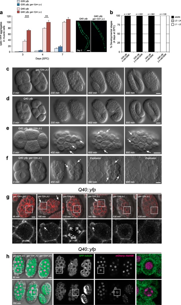
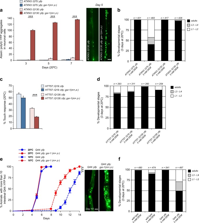
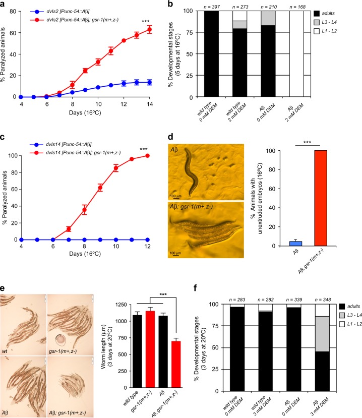
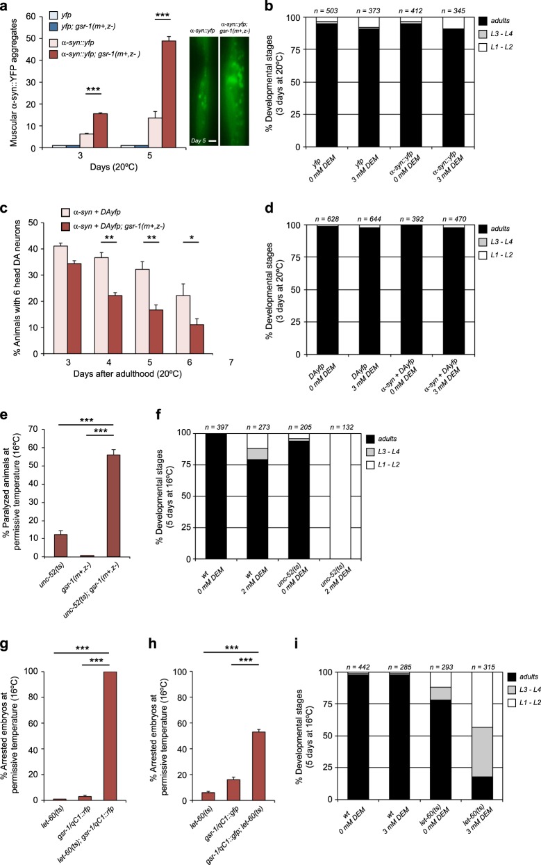
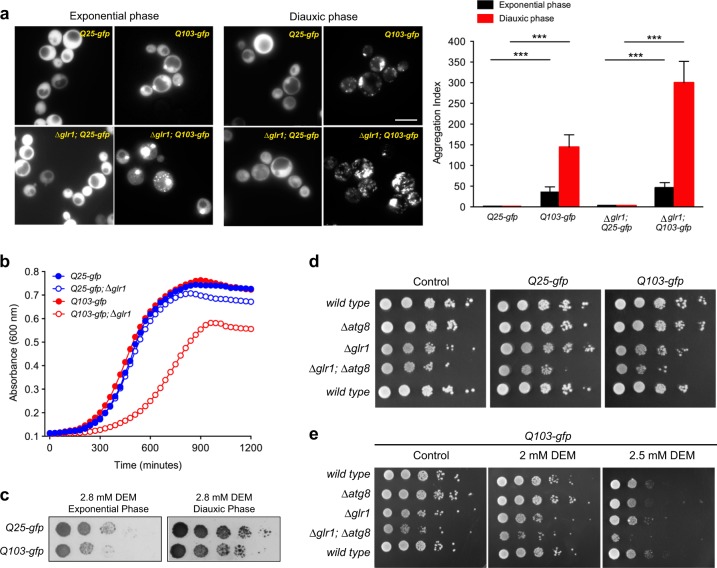
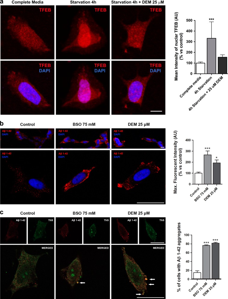
In the presence of aggregation-prone proteins, the cytosol and endoplasmic reticulum (ER) undergo a dramatic shift in their respective redox status, with the cytosol becoming more oxidized and the ER more reducing. However, whether and how changes in the cellular redox status may affect protein aggregation is unknown. Here, we show that C. elegans loss-of-function mutants for the glutathione reductase gsr-1 gene enhance the deleterious phenotypes of heterologous human, as well as endogenous worm aggregation-prone proteins. These effects are phenocopied by the GSH-depleting agent diethyl maleate. Additionally, gsr-1 mutants abolish the nuclear translocation of HLH-30/TFEB transcription factor, a key inducer of autophagy, and strongly impair the degradation of the autophagy substrate p62/SQST-1::GFP, revealing glutathione reductase may have a role in the clearance of protein aggregates by autophagy. Blocking autophagy in gsr-1 worms expressing aggregation-prone proteins results in strong synthetic developmental phenotypes and lethality, supporting the physiological importance of glutathione reductase in the regulation of misfolded protein clearance. Furthermore, impairing redox homeostasis in both yeast and mammalian cells induces toxicity phenotypes associated with protein aggregation. Together, our data reveal that glutathione redox homeostasis may be central to proteostasis maintenance through autophagy regulation.


Conflict of interest statement

The authors declare that they have no conflict of interest.

Figures

Fig. 1
Fig. 1
Phenotypes of the gsr-1 mutation and diethyl maleate (DEM) treatment on C. elegans expressing Q40::YFP protein in muscle cells. a The gsr-1(m+,z-) mutation increases the number of Q40::YFP, but no Q35::YFP, aggregates in worm muscle cells. Data are the mean ± S.E.M. of three independent experiments (n = 10 animals per strain and assay). **p < 0.01; ***p < 0.001 by unpaired, two-tailed Student's t-test. Images show one representative example of muscle Q40::YFP aggregates for each genotype. Scale bar 100 µm. b 2.5 mM DEM treatment causes a fully penetrant L1-L2 larval arrest on worms expressing a Q40::YFP fusion protein in muscle cells while no effect is found in Q35::yfp animals. Data are from three independent experiments (n = total number of animals assayed). c–f Differential interference contrast still images of developing wild-type and gsr-1(m-,z-) embryos (c), Q40::yfp embryos (d) and Q40::yfp; gsr-1(m-,z-) embryos (e, f). Arrows indicate transient blebs. Embryo explosion in (f) is denoted by the sudden disappearance of embryonic cells. The still images in c-f are taken from Movies 1 to 4, respectively. g Control gsr-1(m+,z+) and Q40::yfp; gsr-1(m-,z-) embryos expressing the actin marker LifeAct::mCherry were mounted and recorded together. Bottom row shows higher magnification images of LifeAct::mCherry only. Closed arrows point to examples of membrane blebs whereas double-headed arrows indicate F-actin accumulation at cell vertices. Last frame corresponds to the moment when the eggshell suddenly fills completely and the embryo explodes. The still images are taken from Movie 5. h Control Q40::yfp; gsr-1(m+,z+) and Q40::yfp; gsr-1(m-,z-) embryos expressing the microtubule marker GFP::tubulin and the chromatin marker mCherry::hisH2B (green and magenta in merge, respectively) were mounted and recorded together. Right column shows higher magnification images of boxed area. Strong accumulation of microtubules at the plasma membrane is observed at 60 min (top row). Condensation of chromatin at the nuclear periphery occurs at 350 min. Additional examples are provided in Movie 6. All movie recordings were carried out at 25 °C in H2O except in (h) that was performed in M9 buffer at 25 °C. Time is indicated from start of recording. Scale bar 10 µm
Fig. 2
Fig. 2
Phenotypes of the gsr-1 mutation and diethyl maleate (DEM) treatment on C. elegans expressing polyQ proteins in neurons and intestinal cells. a The gsr-1(m+,z-) mutation increases the number of ATXN3::Q130::YFP aggregates in the worm ventral nerve cord while no effect is found in control worms expressing ATXN3::Q75::YFP fusion protein. Data are the mean ± S.E.M. of three independent experiments (n = 12 animals per strain and assay). ***p < 0.001 by unpaired, two-tailed Student's t-test. Images show one representative example of ventral nerve cord ATXN3::Q130::YFP aggregates for each genotype. Scale bar 20 µm. b 3 mM DEM treatment delays the larval development of worms expressing pan-neuronal ATXN3::Q75::YFP but not ATXN3::Q130::YFP fusion proteins. Data are from three independent experiments (n = total number of animals assayed). c Percentage of touch-responsiveness of wt versus gsr-1(m+,z-) worms expressing HTT57::Q19::CFP and HTT57::Q128::CFP fusion proteins in mechanosensory neurons. Data are the mean ± S.D. of two independent experiments (n ≥ 100 animals per strain and assay). ***p < 0.001 by one-way analysis of variance (ANOVA) with Tukey´s multiple comparison test. d 3 mM DEM treatment does not affect the larval development of worms expressing HTT57::Q19::CFP and HTT57::Q128::CFP fusion proteins in mechanosensory neurons. Data are from three independent experiments (n = total number of animals assayed). e Onset of appearance of YFP fluorescent aggregates in wt versus gsr-1(m+,z-) worms expressing Q44::YFP fusion protein as function of time. Data are the mean ± S.E.M of three independent experiments (n = 50 animals per strain and assay). ***p < 0.001 by log-rank (Mantel–Cox) test. Images show one representative example of intestinal Q44::YFP aggregates for each genotype. Scale bar 50 µm. f 3 mM DEM treatment delays the larval development of worms expressing a Q44::YFP fusion protein in intestinal cells. Data are from three independent experiments (n = total number of animals assayed)
Fig. 3
Fig. 3
Phenotypes of the gsr-1 mutation and diethyl maleate (DEM) treatment on C. elegans expressing the human β-amyloid peptide. a The gsr-1(m+,z-) mutation increases the paralysis of worms expressing the human β-amyloid peptide in muscle cells from the dvIs2 integrated array. Data are the mean ± S.E.M. of three independent experiments (n = 25 animals per strain and assay). ***p < 0.001 by log-rank (Mantel–Cox) test. b 2 mM DEM treatment causes a fully penetrant L1-L2 larval arrest on worms expressing the human β-amyloid peptide in muscle cells. Data are from three independent experiments (n = total number of animals assayed). c When expressing the human β-amyloid peptide in muscle cells from the dvIs14 integrated array, the gsr-1(m+,z-) mutation increases the paralysis of worms much faster than that observed in dvIs2 animals. Data are the mean ± S.E.M. of three independent experiments (n = 25 animals per strain and assay). ***p < 0.001 by log-rank (Mantel–Cox) test. d The gsr-1(m+,z-) mutation provokes a fully penetrant unextruded embryo phenotype in worms expressing the human β-amyloid peptide in muscle cells from the dvIs14 transgene. Data are the mean ± S.E.M. of three independent experiments (n = 50 animals per strain and assay). ***p < 0.001 by unpaired, two-tailed Student's t-test. Images show one representative example of dvIs14 and dvIs14; gsr-1(m+,z-) worms. Scale bar 100 µm. e The gsr-1(m+,z-) mutation delays the development of worms expressing the human β-amyloid peptide in the nervous system. Data are the mean ± S.E.M. of three independent experiments (n = 40 worms per strain and assay). ***p < 0.001 by unpaired, two-tailed Student's t-test. Images show one representative example for each genotype. Scale bar 200 µm. f 3 mM DEM treatment delays the larval development of worms expressing the human β-amyloid peptide in neurons. Data are from three independent experiments (n = total number of animals assayed)
Fig. 4
Fig. 4
Phenotypes of the gsr-1 mutation and diethyl maleate (DEM) treatment on C. elegans expressing human α-synuclein and endogenous metastable proteins. a The gsr-1(m+,z-) mutation increases the number of α-SYN::YFP aggregates in the muscle cells. Aggregate quantification was restricted to aggregates located between the two pharyngeal bulbs. Data are the mean ± S.E.M. of three independent experiments (n = 12 animals per strain and assay). ***p < 0.001 by unpaired, two-tailed Student's t-test. Images show one representative example of muscle α-SYN::YFP aggregates for each genotype. Scale bar 10 µm. b 3 mM DEM treatment does not affect the development of worms expressing the α-SYN::YFP fusion protein in muscle cells. Data are from three independent experiments (n = total number of animals assayed). c The gsr-1(m+,z-) mutation accelerates the age-dependent neurodegeneration of dopaminergic neurons expressing human α-synuclein. Data are the mean ± S.E.M. of three independent experiments (n = 30 animals per strain and assay). *p < 0.05, **p < 0.01 by unpaired, two-tailed Student's t-test. d 3 mM DEM treatment does not affect the development of worms expressing human α-synuclein in dopaminergic neurons. Data are from three independent experiments (n = total number of animals assayed). e The gsr-1(m+,z-) mutation increases the paralysis phenotype of unc-52(e669su250) worms at permissive temperature. Data are the mean ± S.E.M. of three independent experiments (n≥70 animals per strain and assay, scored at day 7 after egg-lay). ***p < 0.001 by unpaired, two-tailed Student's t-test. f 2 mM DEM treatment causes a fully penetrant L1-L2 larval arrest on unc-52(e669su250) worms at permissive temperature. Data are from three independent experiments (n = total number of animals assayed). g, h The gsr-1(m+,z-) mutation increases the embryonic arrest phenotype of let-60(ga89) worms at permissive temperature. g let-60(ga89); gsr-1/qC1::rfp worms produced only arrested embryos while (h) let-60(ga89); gsr-1/qC1::gfp worms only produced both balanced viable progeny and arrested embryos but not unbalanced gsr-1(m+,z-) progeny. Data are the mean ± S.E.M. of three independent experiments (n ≥ 100 embryos/animals per strain and assay). ***p < 0.001 by unpaired, two-tailed Student's t-test. i 3 mM DEM treatment delays the larval development of let-60(ga89) worms at permissive temperature. Data are from three independent experiments (n = total number of animals assayed)
Fig. 5
Fig. 5
The gsr-1 mutation enhances the phenotypes of C. elegans proteostasis models by preventing autophagy function. a The gsr-1(m+,z-) mutation does not modify the diffuse cytoplasmic localization of the HLH-30::GFP reporter under well-fed conditions but strongly inhibits its nuclear translocation after one hour starvation. Data are the mean of three independent experiments (n = 50 animals per strain and assay). ***p < 0.001 by chi2 test. Scale bar 50 µm. b The gsr-1(m+,z-) mutation increases the appearance of SQST-1::GFP foci in embryos generated by parents that have been starved for 24 h. DIC images are provided for embryo position identification. The exposure time for the fluorescence images of sqst-1::gfp embryos was 5x times of that of sqst-1::gfp; gsr-1(m+,z-) embryos. Arrows indicate vacuoles in the sqst-1::gfp; gsr-1(m+,z-) embryos. Scale bar 20 µm. Data are the mean ± S.D. of two independent experiments (n = 50 embryos per strain and assay). ***p < 0.001 by unpaired, two-tailed Student's t-test. c The atg-3(bp412) mutation increases the paralysis of unc-52(e669su250) worms, expressing metastable UNC-52 protein in muscle cells. Data are the mean ± S.E.M. of three independent experiments (n ≥ 50 animals per strain and assay). ***p < 0.001 by log-rank (Mantel–Cox) test of all strains carrying the unc-52(e669su250) allele versus the unc-52 single mutant control. d Expression of the SQST-1::GFP autophagy substrate increases the paralysis onset of unc-52(e669su250) worms and this effect is further enhanced in a gsr-1(m+,z-) background. Data are the mean ± S.E.M. of three independent experiments (n ≥ 50 animals per strain and assay). p < 0.001 by log-rank (Mantel–Cox) test of all strains carrying the unc-52(e669su250) allele versus the unc-52 single mutant control. e Hypodermal seam cells expressing the mCherry::GFP::LGG-1 tandem reporter, imaged at day 1 of adulthood. Autophagosomes (APs) are labeled in yellow while autolysosomes (AL) are labeled in red. Data are the mean ± S.E.M. of four independent experiments (n ≥ 12 animals per strain and assay, with two seam cells quantified for each animal). *p < 0.05, **p < 0.01, ***p < 0.001 by one-way analysis of variance (ANOVA) with Tukey´s multiple comparison test, compared with their respective AP or AL controls. Scale bar 10 µm
Fig. 6
Fig. 6
The protective role of the glutathione system in proteostasis maintenance is conserved in yeast. WT and Δglr1 yeast strains expressing either 25Q-gfp or 103Q-gfp were grown in SC medium. a PolyQ-gfp fusion proteins were visualized by fluorescence microscopy and quantified (aggregation index) in cultures grown to exponential or diauxic phase as described in Materials and methods. The Δglr1 mutation increases the aggregation of 103Q-gfp but not 25Q-gfp. Data are the mean ± S.E.M. of three independent experiments with four biological replicates each. ***p < 0.01 by unpaired, two-tailed Student's t-test. Scale bar 6 µm. b Growth curves were measured in a microplate spectrophotometer for 20 h and only the Δglr1; 103Q-gfp strain shows growth delay. The data are the averages of two independent experiments with three biological replicates each. c Viability of Q25-gfp and Q103-gfp yeast measured by plating serial dilutions (1:10) of cells grown to exponential or diauxic phase in SC plates without or with diethyl maleate (DEM; 2.8 mM). d, e Effect of Δglr1 and/or Δatg8 mutations on the viability of Q25-gfp and Q103-gfp yeast under (d) non-stressed conditions or (e) treated with DEM. Viability was measured by plating serial dilutions (1:10) of cells grown to diauxic phase prior to plating on SC plates
Fig. 7
Fig. 7
The protective role of the glutathione system in proteostasis maintenance is conserved in mammals. a Confocal microscopy images of SH-SY5Y cells in complete medium or starved for 4 h in the absence or presence of DEM (25 µM). Robust nuclear TFEB translocation is found after 4 h of starvation, which is prevented when cells were treated with 25 µM diethyl maleate (DEM). TFEB is shown in red and DAPI staining for nuclei is shown in blue. Data are the mean ± S.E.M. of three independent experiments (n = 15 images with at least 5 cells/image per assay and treatment). ***p < 0.001 by one-way analysis of variance (ANOVA) followed by Newman–Keuls’ test. Scale bar 10 µm. b Confocal microscopy images of SH-SY5Y cells incubated for 18 h in the presence of BSO (75 mM) and DEM (25 µM). A clear increase of intracellular Aβ1–42 staining is found in BSO and DEM-treated cells. Aβ1–42 is shown in red and DAPI staining for nuclei is shown in blue. Data are the mean ± S.E.M. of three independent experiments (n = 45 images with at least 5 cells/image per assay and treatment). *p < 0.01, ***p < 0.001 by one-way ANOVA followed by Newman–Keuls’ test. Scale bar 30 µm. c The Aβ1–42 aggregates in cells treated with BSO and DEM (in red), colocalize with amyloid fibrils labeled by Thioflavin S staining (in green). Colocalization is indicated by white arrows in the merged images. Data are the mean ± S.E.M. of three independent experiments (n ≥ 50 cells per assay and treatment). ***p < 0.001 by one-way ANOVA followed by Newman–Keuls’ test. Scale bar 20 µm

References

    1. Niforou K, Cheimonidou C, Trougakos IP. Molecular chaperones and proteostasis regulation during redox imbalance. Redox Biol. 2014;2:323–32. doi: 10.1016/j.redox.2014.01.017. - DOI - PMC - PubMed
    1. Powers ET, Balch WE. Diversity in the origins of proteostasis networks--a driver for protein function in evolution. Nat Rev Mol Cell Biol. 2013;14:237–48. doi: 10.1038/nrm3542. - DOI - PMC - PubMed
    1. Bayer TA. Proteinopathies, a core concept for understanding and ultimately treating degenerative disorders? Eur Neuropsychopharmacol. 2015;25:713–24. doi: 10.1016/j.euroneuro.2013.03.007. - DOI - PubMed
    1. Pievani M, Filippini N, van den Heuvel MP, Cappa SF, Frisoni GB. Brain connectivity in neurodegenerative diseases--from phenotype to proteinopathy. Nat Rev Neurol. 2014;10:620–33. doi: 10.1038/nrneurol.2014.178. - DOI - PubMed
    1. Go YM, Chandler JD, Jones DP. The cysteine proteome. Free Radic Biol Med. 2015;84:227–45. doi: 10.1016/j.freeradbiomed.2015.03.022. - DOI - PMC - PubMed

Publication types

MeSH terms