Comparative Study
. 2019 Aug;32(4):461-471.
doi: 10.1007/s10334-019-00742-6. Epub 2019 Feb 15.

Detection of axonal degeneration in a mouse model of Huntington's disease: comparison between diffusion tensor imaging and anomalous diffusion metrics

Affiliations
Comparative Study

Detection of axonal degeneration in a mouse model of Huntington's disease: comparison between diffusion tensor imaging and anomalous diffusion metrics

Rodolfo G Gatto et al. MAGMA. 2019 Aug.

Abstract

Objective: The goal of this work is to study the changes in white matter integrity in R6/2, a well-established animal model of Huntington's disease (HD) that are captured by ex vivo diffusion imaging (DTI) using a high field MRI (17.6 T).

Materials and methods: DTI and continuous time random walk (CTRW) models were used to fit changes in the diffusion-weighted signal intensity in the corpus callosum of controls and in R6/2 mice.

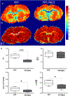
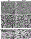
Results: A significant 13% decrease in fractional anisotropy, a 7% increase in axial diffusion, and a 33% increase in radial diffusion were observed between R6/2 and control mice. No change was observed in the CTRW beta parameter, but a significant decrease in the alpha parameter (- 21%) was measured. Histological analysis of the corpus callosum showed a decrease in axonal organization, myelin alterations, and astrogliosis. Electron microscopy studies demonstrated ultrastructural changes in degenerating axons, such as an increase in tortuosity in the R6/2 mice.

Conclusions: DTI and CTRW diffusion models display quantitative changes associated with the microstructural alterations observed in the corpus callosum of the R6/2 mice. The observed increase in the diffusivity and decrease in the alpha CTRW parameter providing support for the use of these diffusion models for non-invasive detection of white matter alterations in HD.

Keywords: Anomalous diffusion; Diffusion tensor imaging; Huntington disease; Magnetic resonance imaging; Mice.


Conflict of interest statement

Conflict of interest Authors of this manuscript have no conflicts of interest to report.

Figures

Fig. 1
Fig. 1
Calculated diffusion tensor imaging (DTI) parameters from control and from Huntington’s disease mouse brains. a Representative T2-weighted images (b = 0) of coronal slices (a′–f′) from six consecutive frontal lobe regions of a mouse showing the four regions of interest (ROIs) centered in the corpus callosum (light blue circles) used in the DTI analysis. b Compiled data from Huntington’s disease (YFP, R6/2) and control (YFP) mice box plots of the fractional anisotropy (FA), axial diffusivity (AD), radial diffusivity (RD), and mean diffusivity (MD), (**p < 0.01), (n = 3 mice per group). Scale bar = 1 mm
Fig. 2
Fig. 2
Calculated anomalous diffusion parameters from control and from Huntington’s disease mouse brains. a Coronal brain slices displaying anomalous diffusion brain maps for α and β/2 fractional order parameters from Huntington’s disease (YFP, R6/2, right) and control (YFP, left) mice. Note the reduced α values in the corpus callosum (CC) of the YFP, R6/2 mice. b Statistical analysis of anomalous diffusion parameters based on selected CC ROIs (circles in β/2 map). Data showed significant differences in Dα,β, α, and 2α/β between the control and diseased animals (*p < 0.05). Note that the most significant differences between both groups were related to the parameter α (***p < 0.001) (n = 3 mice per group). Scale bar = 1 mm
Fig. 3
Fig. 3
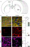
Multi-cellular changes in the corpus callosum of the Huntington’s disease mice reveals increased white matter complexity. a Diagram showing coronal sections and histological regions of interest (ROIs in light blue) centered in the corpus callosum (CC) between the cortex (CCX) and the striatum. b Coronal sections centered in the corpus callosum showing changes in neuronal architecture and axonal orientation by endogenous expression. Yellow fluorescent protein (YFP) can be observed in the R6/2 mice (YFP, R6/2). Note an increase of axonal tortuosity in the R6/2 mice. Nuclear counterstaining with DAPI (Blue). Astrocyte proliferation is labeled by glial fibrillary acid protein (GFAP) and can be observed in white matter (WM) in the HD mice. The amount of myelin basic protein (MBP)—a marker of oligodendrocyte function—is decreased in the HD mice. c Quantitative fluorescence analysis of white matter markers (YFP, GFAP, and MBP) in the corpus callosum of the R6/2 and WT mice (***p < 0.001) (n = 6 mice per group). AU Arbitrary Units. Scale bar = 10 μm
Fig. 4
Fig. 4
Transmission electron microscopy shows complex ultrastructural axonal changes in the corpus callosum of the Huntington’s disease mice R6/2. a Transmission electron microscopy (TEM) from white matter (cross sections) centered in the prefrontal region of the corpus callosum in the wild-type (WT) and Huntington’s mice (R6/2) taken at ×6000 magnification. Scale bar = 10 μm. Note that axonal myelin sheets are stained in black by osmium tetroxide (OsO4). a′ Greater magnification from Fig. 4a (dotted white square) in WT and R6/2 mice shows a decrease in axonal diameters and a relative increase in extra-axonal space and axonal density in the R6/2 mice. Scale bar = 1 μm. b TEM from the corpus callosum WM (longitudinal section) showed an increase in axonal tortuosity (curved double-headed white arrow) compared to similar regions in the control mice (straight white double-headed arrow). IA intra-axonal compartment, EA extra-axonal compartment. Scale bar = 1 μm

Similar articles

Cited by

References

    1. Harper PS (1992) The epidemiology of Huntington’s disease. Hum Genet 89(4):365–376 - PubMed
    1. Pringsheim T, Wiltshire K, Day L, Dykeman J, Steeves T, Jette N (2012) The incidence and prevalence of Huntington’s disease: a systematic review and meta-analysis. Mov Disord 27(9):1083–1091 - PubMed
    1. Walker FO (2007) Huntington’s disease. Lancet 369(9557):218–228 - PubMed
    1. Han I, You Y, Kordower JH, Brady ST, Morfini GA (2010) Differential vulnerability of neurons in Huntington’s disease: the role of cell type-specific features. J Neurochem 113(5):1073–1091 - PMC - PubMed
    1. Perez-Navarro E, Canals JM, Gines S, Alberch J (2006) Cellular and molecular mechanisms involved in the selective vulnerability of striatal projection neurons in Huntington’s disease. Histol Histopathol 21(11):1217–1232 - PubMed

Publication types