

Is contrast-enhanced ultrasound (CEUS) superior to computed tomography angiography (CTA) in detection of endoleaks in post-EVAR patients? A systematic review and meta-analysis
- PMID: 30771104
- PMCID: PMC6430298
- DOI: 10.1007/s40477-019-00364-7
Is contrast-enhanced ultrasound (CEUS) superior to computed tomography angiography (CTA) in detection of endoleaks in post-EVAR patients? A systematic review and meta-analysis
Abstract
Objective: The purpose of this systematic review and meta-analysis was to assess the sensitivity and specificity of contrast-enhanced ultrasound (CEUS) compared to computed tomography angiography (CTA) for the detection of endoleaks within endovascular aortic aneurysm repair (EVAR) surveillance at time of follow up.
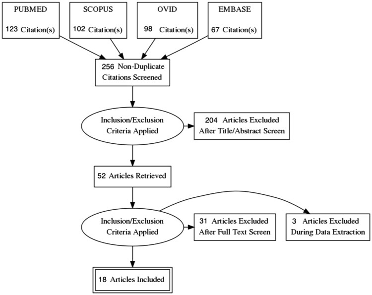
Methods: A comprehensive literature search was undertaken among the four major databases (PubMed, Embase, Scopus and Ovid) to identify all articles assessing diagnostic specificity and accuracy with comparative modality (CEUS vs CTA) for endoleaks in adult patients at time of follow-up following EVAR. Databases where evaluated and assessed to October 2018.
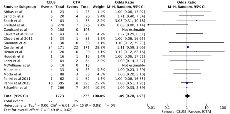
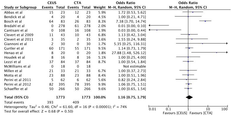
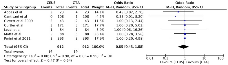
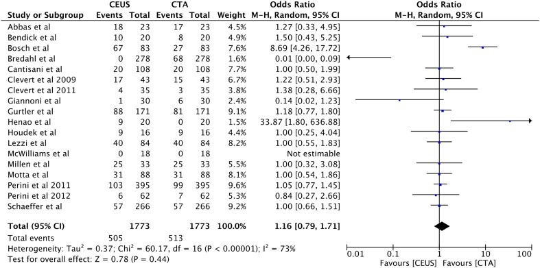
Results: A total of 1773 patients were analysed from across 18 included studies in the quantitative analysis of the parameters of interest. There was no significant difference in detection rate of endoleak type I with detection rate 4.3% for both groups OR 1.09, 95% CI [0.78, 1.53], p = 0.62; type II endoleak detection rate was 22% in the CEUS group vs 23% in the CTA group OR 1.16, 95% CI [0.75-1.79], p = 0.50; while type III detection rate was 1.8% in CEUS group vs 2% in CTA group OR 0.85, 95% CI [0.43, 1.68], p = 0.64. However, the sensitivity rate for endoleak detection was higher in CEUS (p = 0.001) while no difference in specificity rate was noted (p = 0.28). There was higher rate of missed endoleaks in CTA groups (n = 12 vs n = 20).
Conclusion: Evidences from this study suggest that contrast-enhanced ultrasound scan post-EVAR can be utilised as safe and effective method in screening for endoleaks during post-EVAR surveillance without exposing the patient for additional risk of radiation and contrast. CEUS conveys no inferiority to CTA in detecting endoleaks.
Keywords: Angiography; Computerized tomography; Contrast study; EVAR; Ultrasound.
Conflict of interest statement
Conflict of interest
All authors wish to disclose no conflict of interest.
Ethical approval
This article does not contain any direct studies with human participants performed by any of the authors of this paper.
Informed consent
Not applicable.
Figures
Similar articles
-
Can we replace computed tomography angiography by contrast-enhanced ultrasound in the surveillance of patients submitted to aortoiliac aneurysm repair?Vascular. 2022 Aug;30(4):803-808. doi: 10.1177/17085381211027440. Epub 2021 Jun 18. Vascular. 2022. PMID: 34144651
-
Contrast Enhanced Ultrasound can Replace Computed Tomography Angiography for Surveillance After Endovascular Aortic Aneurysm Repair.Eur J Vasc Endovasc Surg. 2016 Dec;52(6):729-734. doi: 10.1016/j.ejvs.2016.07.007. Epub 2016 Oct 17. Eur J Vasc Endovasc Surg. 2016. PMID: 27760698
-
A Systematic Review of Ultrasound or Magnetic Resonance Imaging Compared With Computed Tomography for Endoleak Detection and Aneurysm Diameter Measurement After Endovascular Aneurysm Repair.J Endovasc Ther. 2016 Dec;23(6):936-943. doi: 10.1177/1526602816664878. Epub 2016 Aug 19. J Endovasc Ther. 2016. PMID: 27542700
-
Endovascular Aortic Repair Follow up Protocol Based on Contrast Enhanced Ultrasound Is Safe and Effective.Eur J Vasc Endovasc Surg. 2018 Jul;56(1):40-47. doi: 10.1016/j.ejvs.2018.03.006. Epub 2018 Apr 17. Eur J Vasc Endovasc Surg. 2018. PMID: 29673811
-
Contrast-enhanced ultrasound (CEUS) versus computed tomography angiography (CTA) in detection of endoleaks in post-EVAR patients. Are delayed type II endoleaks being missed? A systematic review and meta-analysis.J Ultrasound. 2015 Jan 17;18(2):91-9. doi: 10.1007/s40477-014-0154-x. eCollection 2015 Jun. J Ultrasound. 2015. PMID: 26191109 Free PMC article. Review.
Cited by
-
Role of Contrast-Enhanced Ultrasound in the Follow-Up after Endovascular Abdominal Aortic Aneurysm Repair.Diagnostics (Basel). 2022 Dec 15;12(12):3173. doi: 10.3390/diagnostics12123173. Diagnostics (Basel). 2022. PMID: 36553180 Free PMC article.
-
Novel Use of Contrast-enhanced Ultrasound in the Pretreatment Planning Prior to Endovascular Repair of Endoleak after Endovascular Aortic Aneurysm Repair in a Patient with Chronic Renal Insufficiency: A Case Report and Literature Review.J Med Ultrasound. 2021 Apr 23;30(1):54-58. doi: 10.4103/JMU.JMU_173_20. eCollection 2022 Jan-Mar. J Med Ultrasound. 2021. PMID: 35465604 Free PMC article.
-
EVAR Follow-Up with Ultrasound Superb Microvascular Imaging (SMI) Compared to CEUS and CT Angiography for Detection of Type II Endoleak.Diagnostics (Basel). 2022 Feb 18;12(2):526. doi: 10.3390/diagnostics12020526. Diagnostics (Basel). 2022. PMID: 35204615 Free PMC article.
-
Multi-Energy Low-Kiloelectron Volt versus Single-Energy Low-Kilovolt Images for Endoleak Detection at CT Angiography of the Aorta.Radiol Cardiothorac Imaging. 2024 Apr;6(2):e230217. doi: 10.1148/ryct.230217. Radiol Cardiothorac Imaging. 2024. PMID: 38451189 Free PMC article. Clinical Trial.
-
Diagnostic accuracy of microvascular flow imaging ultrasound for endoleak detection after endovascular aortic aneurysm repair: a systematic review and meta-analysis.Pol J Radiol. 2024 Aug 22;89:e414-e419. doi: 10.5114/pjr/190502. eCollection 2024. Pol J Radiol. 2024. PMID: 39257925 Free PMC article. Review.
References
-
- Choke E, Thompson M. Endoleak after endovascular aneurysm repair: current concepts. J Cardiovasc Surg. 2004;45:349. - PubMed
Publication types
MeSH terms
Substances
LinkOut - more resources
Full Text Sources
