. 2019 May 1:449:66-75.
doi: 10.1016/j.canlet.2019.02.009. Epub 2019 Feb 13.

Squalamine blocks tumor-associated angiogenesis and growth of human breast cancer cells with or without HER-2/neu overexpression

Affiliations

Squalamine blocks tumor-associated angiogenesis and growth of human breast cancer cells with or without HER-2/neu overexpression

Diana C Márquez-Garbán et al. Cancer Lett. .

Abstract

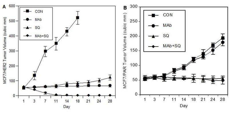
Angiogenesis is critical for breast cancer progression. Overexpression of HER-2/neu receptors occur in 25-30% of breast cancers, and treatment with trastuzumab inhibits HER-2-overexpressing tumor growth. Notably, HER-2-mediated signaling enhances vascular endothelial growth factor (VEGF) secretion to increase tumor-associated angiogenesis. Squalamine (aminosterol compound) suppresses VEGF-induced activation of kinases in vascular endothelial cells and inhibits tumor-associated angiogenesis. We assessed antitumor effects of squalamine either alone or with trastuzumab in nude mice bearing breast tumor xenografts without (MCF-7) or with HER2-overexpression (MCF-7/HER-2). Squalamine alone inhibited progression of MCF-7 tumors lacking HER2 overexpression, and squalamine combined with trastuzumab elicited marked inhibition of MCF-7/HER2 growth exceeding that of trastuzumab alone. MCF-7/HER-2 cells secrete higher levels of VEGF than MCF-7 cells, but squalamine elicited no growth inhibition of either MCF-7/HER-2 or MCF-7 cells in vitro. However, squalamine did stop growth of human umbilical vein endothelial cells (HUVECs) and reduced VEGF-induced endothelial tube-like formations in vitro. These effects correlated with blockade of focal adhesion kinase phosphorylation and stress fiber assembly in HUVECs. Thus, squalamine effectively inhibits growth of breast cancers with or without HER-2-overexpression, an effect due in part to blockade of tumor-associated angiogenesis.

Keywords: Breast cancer; MCF-7; Squalamine; Trastuzumab; Tumor-associated angiogenesis; VEGF.


Conflict of interest statement

Conflict of interest

Richard J. Pietras has consulted with Astra-Zeneca, Pfizer and Genentech. The remaining authors declare no conflicts of interest.

Figures

Figure 1.
Figure 1.
Squalamine inhibits growth of human breast tumor xenografts in vivo. A) Squalamine inhibits growth of MCF-7/HER2 breast tumor xenografts in vivo and enhances antitumor effects of trastuzumab. MCF-7/HER-2-overexpressing breast cancer cells were subcutaneously inoculated in nude mice. After 7 days, animals with tumors of comparable size were randomized to treatment with trastuzumab (Mab; 8 mg/kg loading dose followed by a 4 mg/kg dose administrated weekly thereafter), squalamine (SQ; 2 mg/kg on days 1-28), or trastuzumab administrated in combination with squalamine (Mab+SQ). The control group (CON) received appropriate vehicle injections on days 1-28. Animals with tumors exceeding 500-mm3 were sacrificed as per our approved institutional animal research committee protocol. After day 28, the experiment was terminated. Results are expressed as mean ± SEM. Antitumor effects of MAb and SQ alone groups were significantly different from those of the CON group; and antitumor effects of the MAb+SQ combination treatment group were significantly different from those of CON and from either MAb or SQ treatments as single agents (all at P < 0.01). B) Squalamine inhibits growth of MCF-7 breast tumor xenografts but does not enhance effects of trastuzumab in tumors without HER2 overexpression. MCF-7 cells were inoculated subcutaneously as above, and animals with tumors of comparable size were randomized to treatment with control (CON), trastuzumab (MAb), squalamine (SQ) or a combination of these two agents (MAb+SQ). The control group received appropriate vehicle injections during the course of the study as in A) above. After day 28, the experiment was terminated, with results expressed as mean ± SEM. Antitumor effects of SQ alone or MAb+SQ groups were both significantly different from those of the CON group (P<0.001); but antitumor effects of the MAb treatment alone as expected were not significantly different from those of the CON group.
Figure 2.
Figure 2.
Immunohistochemical staining for vascular endothelial cells in MCF-7/HER-2 xenograft tissues obtained from nude mice treated for seven days with either squalamine (SQ) or trastuzumab (MAb) administered alone or in combination (MAb/SQ). Microvessels were stained using a polyclonal antibody against vWf and were randomly counted from 5 different high-power fields (×200) per mouse tumor. The values are represented as means ± SEM of microvessel density per high-power field (n = 3). Treatment with squalamine (SQ) alone or a combination of squalamine with trastuzumab (MAb/SQ) elicited a significant reduction in microvessel density as compared to controls (CON) or to trastuzumab (MAb) alone (P < 0.01, by student’s t-test).
Figure 3.
Figure 3.
VEGF secretion by MCF-7 breast cancer cells with or without HER-2/neu gene overexpression. A) Cells were grown in vitro, either with control medium (Con) or medium containing 1.6 μM squalamine (SQ). After 48 hours, media were collected, and VEGF levels were measured by ELISA assay. Data are represented as mean ± SEM. VEGF levels secreted by MCF-7/HER2 (MCF/HER2) cells were significantly different than those secreted by MCF-7 parental cells without HER2-overexpression (MCF/PAR) when treated with either control or squalamine (P < 0.05). Addition of squalamine did not affect the level of VEGF secretion by either MCF/PAR or MCF/HER2 cells. B) Western immunoblot showing VEGF expression levels after cells were treated with varying doses of squalamine. MCF7/PAR and MCF7/HER2 cells were grown in vitro and treated with increasing doses of squalamine. After 48 hours, the supernatant was collected and concentrated as described in methods. Western immunoblots were done using anti-VEGF antibody (Thermo Fisher). The results indicate no difference in the expression levels of VEGF secreted into the extracellular medium after squalamine treatment.
Figure 4.
Figure 4.
In vitro HUVEC and MCF-7/HER-2 cell proliferation. a) Squalamine inhibits VEGF-induced proliferation of endothelial cells in vitro. HUVECs were grown in the presence of VEGF (50 ng/ml), squalamine (SQ; 3.2 μM), combinations of VEGF with squalamine (VEGF/SQ 3.2 μM ; VEGF/SQ 1.6 μM; VEGF /SQ 0.16 μ M and VEGF/SQ 0.016 μM), or control medium alone (Con). Results show that VEGF alone, but not SQ alone, stimulates HUVEC proliferation by day 8 (P<0.001); while SQ elicits a dose-dependent reduction in VEGF-induced HUVEC proliferation at doses ranging from 0.16 to 3.2 μM by day 8 (P<0.01). b) Squalamine does not directly affect growth of MCF-7/HER-2 breast cancer cells in vitro. Results show that cell numbers of MCF-7/HER2 cells are unchanged by incubation with either VEGF or SQ as compared to controls (CON) (P>0.05). All data are from duplicate determinations of cell numbers and the values are represented as mean ± SEM from 3 independent experiments.
Figure 5.
Figure 5.
Regression of VEGF-induced HUVEC tube-like structures after squalamine treatment. HUVECs were cultured in serum-free medium on the surface of Geltrex with the indicated treatments. A) vehicle (CONTROL); B) squalamine 1 μM (SQUAL); C) VEGF 50 ng/ml; D) VEGF (50 ng/ml) + squalamine (1 μM) (SQ+VEGF). After 18 h incubation, endothelial cell tubular structures were observed under a fluorescent microscope and photographed with a 20× objective (top panels) and analyzed by Angiogenesis Analyzer ImageJ as described in methods. Middle and lower panels show corresponding skeletons of tubular networks identifying master segments (orange), meshes (blue sky), master junctions (red), branches (green), nodes (red surrounded by blue) and segments (magenta). E) and F) ImageJ analyzed parameters of tube formation E) node number, F) number of master segments, meshes and master junctions were calculated. Values are the mean and SEM calculated from triplicates in 3 independent experiments (n=3, *p<0.05; **p<0.01). HUVEC cells in those plates in which the networked capillary tubes were more effectively inhibited by squalamine showed marked alteration in their shape and size, in contrast with the more characteristic spindle-shaped cells that form capillary-like tubes either in the absence of squalamine or in the presence of very low doses of squalamine.
Figure 6.
Figure 6.
Squalamine blocks FAK phosphorylation and formation of actin stress fibers in HUVECs exposed to VEGF. a) HUVECs were made quiescent and either treated with a) control (CON); or b) treated with squalamine (SQ; 3.2 μ M) alone for 60 min; c) treated with 50 ng/ml VEGF for 10 min; and compared with d) cells pre-treated with squalamine (3.2 μ M) for 60 min and 50 ng/ml VEGF for 10 min. FAK phosphorylation (green signal) was detected using a polyclonal antibody anti-FAK [pY397]; and F-actin (red signal) was detected using rhodamine-conjugated phalloidin. Cells were examined by confocal microscopy and DAPI was used for nuclear staining (blue). Results show that VEGF stimulates FAK phosphorylation (c1) as compared to control (a1) and squalamine (b1) treatment, while this effect is inhibited by combined treatment with squalamine (d1). Further, VEGF also induces a reorganization of actin stress fibers (c2) as compared to control (a2) and squalamine (b2) treatment, while this action is reversed by dual treatment with squalamine (d2). The lower panel (a3-d3) presents an overlay of the green and red signals for FAK and F-actin, respectively for each of the treatment groups. The bulk of HUVECs responded to VEGF-induced FAK phosphorylation as reported by others [78] as well as to squalamine-dependent disruption of VEGF-induced FAK phosphorylation. Representative fields are shown, based on data obtained in 5 different experiments.
Figure 7.
Figure 7.
Squalamine blocks FAK phosphorylation in HUVECs exposed to VEGF. HUVECs were incubated in the presence of VEGF, squalamine or a combination of both VEGF and squalamine A) HUVEC cells were treated with control vehicle (VH), VEGF (50 ng/ml), squalamine (SQ; 1 μM) or VEGF in combination with squalamine (VEGF+SQ). Lysates were prepared and processed as described in materials and methods. Western blotting was done with monoclonal antibodies against phosphoFAK (upper panel) and total FAK (lower panel). B) HUVEC cells were treated with control vehicle (VH) or increasing concentrations of squalamine from 0.001- 1 μM for 1 hour in vitro. Immunoblotting was done with anti-phospho (Y397) FAK. For loading control, membranes were stripped and reprobed with anti total FAK.

Similar articles

Cited by

References

    1. Coussens L, Yang-Feng TL, Liao YC, Chen E, Gray A, McGrath J, Seeburg PH, Libermann TA, Schlessinger J, Francke U, et al., Tyrosine kinase receptor with extensive homology to EGF receptor shares chromosomal location with neu oncogene, Science 230(4730) (1985) 1132–1139. - PubMed
    1. Semba K, Kamata N, Toyoshima K, Yamamoto T, A v-erbB-related protooncogene, c-erbB-2, is distinct from the c-erbB-1/epidermal growth factor-receptor gene and is amplified in a human salivary gland adenocarcinoma, Proc Natl Acad Sci U S A 82(19) (1985) 6497–6501. - PMC - PubMed
    1. Harris JR, Lippman ME, Veronesi U, Willett W, Breast cancer (3), N Engl J Med 327(7) (1992) 473–480. - PubMed
    1. Lemoine NR, Staddon S, Dickson C, Barnes DM, Gullick WJ, Absence of activating transmembrane mutations in the c-erbB-2 proto-oncogene in human breast cancer, Oncogene 5(2) (1990) 237–239. - PubMed
    1. Press MF, Slamon DJ, Flom KJ, Park J, Zhou JY, Bernstein L, Evaluation of HER-2/neu gene amplification and overexpression: comparison of frequently used assay methods in a molecularly characterized cohort of breast cancer specimens, J Clin Oncol 20(14) (2002) 3095–3105. - PubMed

Publication types

MeSH terms