

Nitro-fatty acids protect against steatosis and fibrosis during development of nonalcoholic fatty liver disease in mice
- PMID: 30772307
- PMCID: PMC6444056
- DOI: 10.1016/j.ebiom.2019.02.019
Nitro-fatty acids protect against steatosis and fibrosis during development of nonalcoholic fatty liver disease in mice
Abstract
Background: Nonalcoholic fatty liver disease (NAFLD) and resulting nonalcoholic steatohepatitis (NASH) are reaching global epidemic proportions. Lack of non-invasive diagnostic tools and effective therapies constitute two of the major hurdles for a bona fide treatment and a reversal of NASH progression and/or regression of the disease. Nitro-oleic acid (OA-NO2) has been proven effective in multiple experimental models of inflammation and fibrosis. Thus, the potential benefit of in vivo administration of OA-NO2 to treat advanced NAFLD was tested herein in a model of long-term NASH diet-induced liver damage.
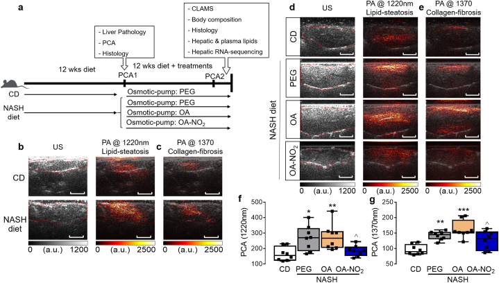
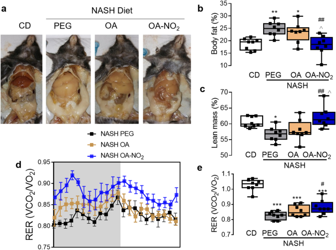
Methods: Non-invasive imaging (e.g. photoacustic-ultrasound (PA-US)) was pursued to establish advanced experimental model of NASH in mice in which both steatosis and fibrosis were diagnosed prior experimental therapy with OA-NO2. Experimental controls included equimolar amounts of the non-nitrated oleic acid (OA). CLAMS and NMR-based analysis was used for energy metabolism.
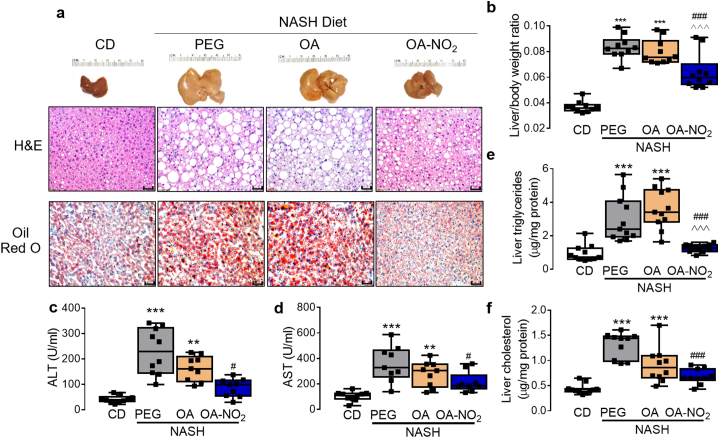
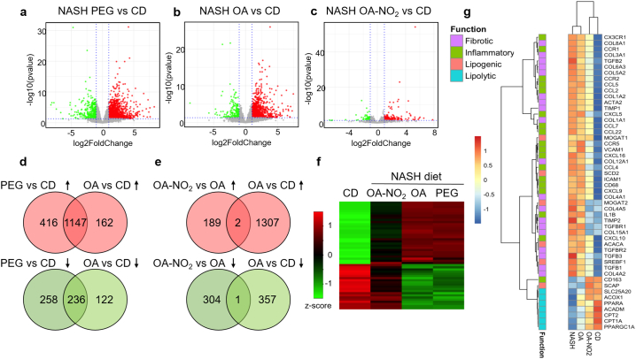
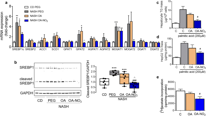
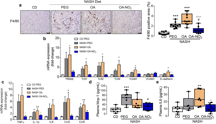
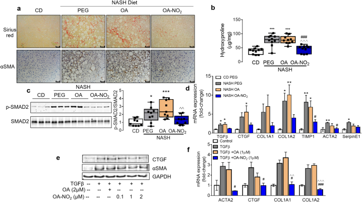
Findings: CLAMS and NMR-based analysis demonstrates that OA-NO2 improves body composition and energy metabolism and inhibits hepatic triglyceride (TG) accumulation. Photoacoustic-ultrasound imaging revealed a robust inhibition of liver steatosis and fibrosis by OA-NO2. RNA-sequencing analysis uncovered inflammation and fibrosis as major pathways suppressed by OA-NO2 administration, as well as regulation of lipogenesis and lipolysis pathways, with a robust inhibition of SREBP1 proteolytic activation and subsequent lipogenesis gene expression by OA-NO2. These results were further supported by histological analysis and quantification of lipid accumulation, lobular inflammation (F4/80 staining) and fibrosis (collagen deposition, αSMA staining) as well as established parameters of liver damage (ALT). In vitro studies indicate that OA-NO2 inhibits TG biosynthesis and accumulation in hepatocytes and inhibits fibrogenesis in human stellate cells.
Interpretation: OA-NO2 improve steatohepatitis and fibrosis and may constitute an effective therapeutic approach against advanced NAFLD that warrants further clinical evaluation.
Keywords: Nitro-fatty acids; Non-alcoholic Steatohepatitis; Non-alcoholic fatty liver disease; Non-invasive liver imaging, liver fibrosis.
Copyright © 2019. Published by Elsevier B.V.
Figures
Comment in
-
Nitro-oleic acid as a new drug candidate for non-alcoholic steatohepatitis.EBioMedicine. 2019 Apr;42:32-33. doi: 10.1016/j.ebiom.2019.03.059. Epub 2019 Apr 1. EBioMedicine. 2019. PMID: 30948353 Free PMC article. No abstract available.
Similar articles
-
Calycosin attenuates triglyceride accumulation and hepatic fibrosis in murine model of non-alcoholic steatohepatitis via activating farnesoid X receptor.Phytomedicine. 2017 Feb 15;25:83-92. doi: 10.1016/j.phymed.2016.12.006. Epub 2016 Dec 13. Phytomedicine. 2017. PMID: 28190475
-
Amelioration by chicory seed extract of diabetes- and oleic acid-induced non-alcoholic fatty liver disease (NAFLD)/non-alcoholic steatohepatitis (NASH) via modulation of PPARα and SREBP-1.Food Chem Toxicol. 2013 Aug;58:198-209. doi: 10.1016/j.fct.2013.04.018. Epub 2013 Apr 18. Food Chem Toxicol. 2013. PMID: 23603006
-
Tcf7l2 in hepatocytes regulates de novo lipogenesis in diet-induced non-alcoholic fatty liver disease in mice.Diabetologia. 2023 May;66(5):931-954. doi: 10.1007/s00125-023-05878-8. Epub 2023 Feb 10. Diabetologia. 2023. PMID: 36759348 Free PMC article.
-
Emerging and Established Therapeutic Approaches for Nonalcoholic Fatty Liver Disease.Clin Ther. 2021 Sep;43(9):1476-1504. doi: 10.1016/j.clinthera.2021.07.013. Epub 2021 Aug 24. Clin Ther. 2021. PMID: 34446271 Review.
-
SREBP Regulation of Lipid Metabolism in Liver Disease, and Therapeutic Strategies.Biomedicines. 2023 Dec 12;11(12):3280. doi: 10.3390/biomedicines11123280. Biomedicines. 2023. PMID: 38137501 Free PMC article. Review.
Cited by
-
Niche preclinical and clinical applications of photoacoustic imaging with endogenous contrast.Photoacoustics. 2023 Jul 17;32:100533. doi: 10.1016/j.pacs.2023.100533. eCollection 2023 Aug. Photoacoustics. 2023. PMID: 37636547 Free PMC article. Review.
-
The Emerging Therapeutic Potential of Nitro Fatty Acids and Other Michael Acceptor-Containing Drugs for the Treatment of Inflammation and Cancer.Front Pharmacol. 2020 Sep 3;11:1297. doi: 10.3389/fphar.2020.01297. eCollection 2020. Front Pharmacol. 2020. PMID: 33013366 Free PMC article. Review.
-
Nitro Fatty Acids (NO2-FAs): An Emerging Class of Bioactive Fatty Acids.Molecules. 2021 Dec 13;26(24):7536. doi: 10.3390/molecules26247536. Molecules. 2021. PMID: 34946618 Free PMC article. Review.
-
Dysregulated oxalate metabolism is a driver and therapeutic target in atherosclerosis.Cell Rep. 2021 Jul 27;36(4):109420. doi: 10.1016/j.celrep.2021.109420. Cell Rep. 2021. PMID: 34320345 Free PMC article.
-
Recent advancements in molecular photoacoustic tomography.JPhys Photonics. 2025 Jul 31;7(3):032003. doi: 10.1088/2515-7647/adf167. Epub 2025 Jul 28. JPhys Photonics. 2025. PMID: 40734710 Free PMC article. Review.
References
-
- Haas J.T., Francque S., Staels B. Pathophysiology and mechanisms of nonalcoholic fatty liver disease. Annu Rev Physiol. 2016;78:181–205. - PubMed
-
- Younossi Z., Anstee Q.M., Marietti M., Hardy T., Henry L., Eslam M. Global burden of NAFLD and NASH: trends, predictions, risk factors and prevention. Nat Rev Gastroenterol Hepatol. 2018;15(1):11–20. - PubMed
-
- Younossi Z.M., Koenig A.B., Abdelatif D., Fazel Y., Henry L., Wymer M. Global epidemiology of nonalcoholic fatty liver disease-meta-analytic assessment of prevalence, incidence, and outcomes. Hepatology. 2016;64(1):73–84. - PubMed
-
- Rinella M.E., Sanyal A.J. Management of NAFLD: a stage-based approach. Nat Rev Gastroenterol Hepatol. 2016;13(4):196–205. - PubMed
MeSH terms
Substances
Grants and funding
LinkOut - more resources
Full Text Sources
Medical
Molecular Biology Databases
Miscellaneous
