Review
. 2019 May 1:487:66-74.
doi: 10.1016/j.mce.2019.02.005. Epub 2019 Feb 14.

Relaxin and fibrosis: Emerging targets, challenges, and future directions

Affiliations
Review

Relaxin and fibrosis: Emerging targets, challenges, and future directions

Anthony J Kanai et al. Mol Cell Endocrinol. .

Abstract

The peptide hormone relaxin is well-known for its anti-fibrotic actions in several organs, particularly from numerous studies conducted in animals. Acting through its cognate G protein-coupled receptor, relaxin family peptide receptor 1 (RXFP1), serelaxin (recombinant human relaxin) has been shown to consistently inhibit the excessive extracellular matrix production (fibrosis) that results from the aberrant wound-healing response to tissue injury and/or chronic inflammation, and at multiple levels. Furthermore, it can reduce established scarring by promoting the degradation of aberrant extracellular matrix components. Following on from the review that describes the mechanisms and signaling pathways associated with the extracellular matrix remodeling effects of serelaxin (Ng et al., 2019), this review focuses on newly identified tissue targets of serelaxin therapy in fibrosis, and the limitations associated with (se)relaxin research.

Keywords: Challenges and future directions; Emerging targets; Extracellular matrix; Fibrosis; RXFP1; Relaxin.


Conflict of interest statement

Conflict of interest statement: The authors report no conflict of interest.

Figures

Figure 1.
Figure 1.
Bladder cystometrograms (CMGs) and external urethral sphincter (EUS) electromyograms (EMGs) from irradiated mice with and without serelaxin treatment. A, Method for selective irradiation of the urinary bladder. B‐E, CMGs/EUS‐EMGs in decerebrated mice. B, Control, nonirradiated mouse (N = 6). C, Nonirradiated mouse treated with serelaxin (400 μg/kg/day) for 2 weeks (N = 2). D, Irradiated mouse with saline infusion via a subcutaneous osmotic pump for 2 weeks (N = 6). E, Irradiated mouse with serelaxin infusion (400 μg/kg/day) via a subcutaneous osmotic pump for 2 weeks (N = 4). Treatment in D and E commenced 7 weeks after irradiation. Serelaxin treated mice exhibited more efficient voiding, longer intercontractile intervals, higher bladder compliances, and a normalized EUS activity. Used with permission from (Ikeda et al., 2018).
Figure 2.
Figure 2.
Passive properties, bladder wall compliance, detrusor contractility and collagen content changes in chronic radiation cystitis and its reversal by serelaxin treatment. A, The bladders were isolated at 1, 2, 4, 6, and 9 weeks post‐irradiation and contractile function was measured in organ bath experiments (N = 3 each time point). Passive tension profiles (an indicator of tissue stiffness) showed significant increases at 6‐9 weeks post‐irradiation. B, Passive tension recorded in Ca2+‐free Tyrode’s solution demonstrated that serelaxin decreased tension generation, compared to saline treated irradiated mice, suggesting that this effect was due to changes in the elastic properties of the bladder and not smooth muscle relaxation, (N = 5 each). C and D, At 9 weeks post‐irradiation, mouse bladders showed increased passive tension and decreased active force generation (red traces) compared to nonirradiated mice (green traces). Two weeks treatment with serelaxin (subcutaneous, 400 μg/kg/day) commenced 7‐week post‐ irradiation resulted in a passive tension profile similar to nonirradiated controls and increased contractile responses to EFS (blue traces and bars, N = 4 for control and N = 5 each for irradiated + vehicle or serelaxin). E, Force generation in response to EFS were enhanced in serelaxin‐treated compared to vehicle treated irradiated mouse bladders (*P < 0.05, Wilcoxon ranked sum test). Responses to muscarinic agonist, oxotremorine‐M and 120 mM KCl were not significantly different between the groups (N = 4 for control and N = 5 each for irradiated + vehicle or serelaxin). F, The expression of L‐type Ca2+channels (Cav1.2) was decreased in the detrusor layer of mice with chronic radiation cystitis and was increased following serelaxin treatment (N = 3 each). G, Van Gieson staining of control mouse bladder sections. H, Sections from irradiated bladders showed denuding of the UT and significant collagen staining in the lamina propria (LP) and throughout the detrusor. I, Mice treated with serelaxin showed a decrease in bladder collagen content that was comparable to nonirradiated mice and an intact urothelial layer. J, Collagen:tissue ratio was analyzed using ImageJ (N = 4 each group, Wilcoxon ranked sum test, * indicate P < 0.01 vs non‐irradiated control and **P < 0.01 vs irradiated + vehicle). Used with permission from (Ikeda et al., 2018).
Figure 3.
Figure 3.
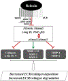
Summary of the mechanisms of relaxin’s anti-fibrotic actions that are mediated through RXFP1 receptors and RXFP1–AT2 receptor heterodimers. Relaxin specifically ameliorates the effects of pro-fibrotic stimuli such as TGF-β1 and Ang II, the former by inhibiting Smad2 (pSmad2) and/or Smad3 (pSmad3) phosphorylation, which is dependent at least in part on the pERK½, NO and Notch-1 pathways. This causes decreased expression and deposition of interstitial (types I, III and V) and basement membrane (type IV) collagens and reduced activity of TIMP-1 and TIMP-2, accompanied by increased expression and activity of various MMPs, includingMMP-1/13, MMP-2 and/or MMP-9. The end result is a decrease in the rate of collagen deposition and increased collagen degradation, allowing clearance of the fibrotic scar.
Figure 4.
Figure 4.
Immunohistological and immunocytochemical staining of tissues and cells for serelaxin and hormone receptors. A. Aminoethylcarbazole staining (red) of relaxin binding to the volar oblique ligament, counterstained with hematoxylin (magnification, x100; see ref 20). B. Ligament staining. Relaxin binding to an ACL specimen from an 81-year-old woman. C. Relaxin staining to an ACL specimen from a 15-year-old girl (for B and C: original magnification, × 100. Positive regions appear red. See ref 21). D. RXFP1 staining in VOL fibroblasts. Cells from a ligament explant culture were fixed and stained with rabbit anti-human RXFP1 coupled to fluorescein (magnification, ×1000; see (Cooney et al., 2009)). E. RXFP1 expression in basement mem-brane from a labial skin biopsy. The image shows localization of rabbit anti-human RXFP1 antibodies (dark) to cells that rest on the basement membrane in a labial specimen from a pediatric patient (magnification, ×400). Images used with permission from (Cooney et al., 2009; Galey et al., 2003; Lubahn et al., 2006).
Figure 5.
Figure 5.
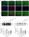
Changes of α-SMA expression after treatment with serelaxin. (A) Immunostaining of α-SMA. Bar = 100 μm. (B) Expression of α-SMA mRNA assessed with RT-PCR. (C) The statistical result of α-SMA mRNA expression. (D) Expression of α-SMA assessed with Western blot. (E) The statistical result of α-SMA expression. Serelaxin inhibits α-SMA expression in TGF-β1-induced cells. *p < 0.05 versus control group, #p < 0.05 versus TGF-β1 group. Used with permission from (Wu et al., 2018).

References

    1. Aitken KJ, Bagli DJ, 2009. The bladder extracellular matrix. Part I: architecture, development and disease. Nat. Rev. Urol 6, 596–611. - PubMed
    1. Bathgate RA, Halls ML, van der Westhuizen ET, Callander GE, Kocan M, Summers RJ, 2013. Relaxin family peptides and their receptors. Physiol. Rev 93, 405–480. - PubMed
    1. Baues M, Dasgupta A, Ehling J, Prakash J, Boor P, Tacke F, Kiessling F, Lammers T, 2017. Fibrosis imaging: Current concepts and future directions. Adv. Drug Deliv. Rev 121, 9–26. - PMC - PubMed
    1. Bennett RG, 2009. Relaxin and its role in the development and treatment of fibrosis. Transl. Res 154, 1–6. - PMC - PubMed
    1. Boyle PM, Hakim JB, Zahid S, Franceschi WH, Murphy MJ, Prakosa A, Aronis KN, Zghaib T, Balouch M, Ipek EG, Chrispin J, Berger RD, Ashikaga H, Marine JE, Calkins H, Nazarian S, Spragg DD, Trayanova NA, 2018. The fibrotic substrate in persistent atrial fibrillation patients: comparison between predictions from computational modeling and measurements from focal impulse and rotor mapping. Front. Physiol 9, 1151. - PMC - PubMed

Publication types

LinkOut - more resources