

Case Reports
doi: 10.1080/01652176.2018.1535216.
A minimally invasive partial condylectomy and temporal bone resection for the treatment of a suspected chronic synovial sepsis of the temporomandibular joint in a 3.5-year-old paint horse gelding
Affiliations
- PMID: 30773124
- PMCID: PMC6830993
- DOI: 10.1080/01652176.2018.1535216
Item in Clipboard
Case Reports
A minimally invasive partial condylectomy and temporal bone resection for the treatment of a suspected chronic synovial sepsis of the temporomandibular joint in a 3.5-year-old paint horse gelding
Vet Q.
2018 Dec.
No abstract available
Keywords: Equine; arthroscopy; horse; mandibular condylectomy; septic arthritis; temporomandibular joint.
Figures

Fluctuant swelling at the level of the right TMJ (pointed by black arrow).
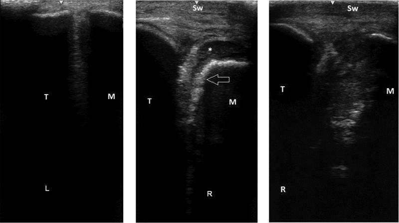
a) Ultrasonogram of the lateral aspect of the unaffected left TMJ. b) Preoperative ultrasonogram of the affected right TMJ; with clear soft tissue swelling (Sw), showing obvious irregular articular mandibular bone margins (Arrow) and a well-defined hypoechoic area dorsal and dorsolateral to it (*). c) Postoperative ultrasonogram, 6 weeks after arthroscopic debridement, showing a spacious right TMJ. T: Temporal bone, M: Mandible, L: Left TMJ, R: Right TMJ.

a) Intra-operative arthroscopic image, debridement of devitalized bone with a fenestrated, 3 mm, ethmoid arthroscopic rongeur (Sontec Instruments). b) debridement until healthy subchondral bone was reached.

a) Transverse CT image at the level of the temporomandibular joint; right on the right side. Partial condylectomy of the mandibular condyle (white arrows) is seen. Note the disuse osteopenia of the right mandible compared to the left. b) 3-D CT image of the right temporo-mandibular joint. Note the irregular contour defect due to the partial condylectomy of the mandibular condyle.
Similar articles
-
Mandibular condylectomy and meniscectomy for the treatment of septic temporomandibular joint arthritis in a horse.Vet Surg. 2006 Oct;35(7):663-8. doi: 10.1111/j.1532-950X.2006.00205.x. Vet Surg. 2006. PMID: 17026552
-
Arthroscopic treatment of temporomandibular joint sepsis in a horse.Vet Surg. 2005 Jan-Feb;34(1):55-8. doi: 10.1111/j.1532-950x.2005.00010.x. Vet Surg. 2005. PMID: 15720597
-
Mandibular condylectomy in a horse.J Am Vet Med Assoc. 1989 Jul 1;195(1):101-2. J Am Vet Med Assoc. 1989. PMID: 2759880
-
[Temporomandibular joint (TMJ): Condyle hyperplasia and condylectomy].Rev Stomatol Chir Maxillofac Chir Orale. 2016 Sep;117(4):259-65. doi: 10.1016/j.revsto.2016.07.021. Epub 2016 Aug 23. Rev Stomatol Chir Maxillofac Chir Orale. 2016. PMID: 27567190 Review. French.
-
Associated bony procedures for preservation.Atlas Oral Maxillofac Surg Clin North Am. 1996 Sep;4(2):107-17. Atlas Oral Maxillofac Surg Clin North Am. 1996. PMID: 11873690 Review. No abstract available.
Cited by
-
Long-Term Outcome of Horses Undergoing Unilateral Mandibular Condylectomy and Meniscectomy for Temporomandibular Joint Disease.Front Vet Sci. 2022 May 2;9:898096. doi: 10.3389/fvets.2022.898096. eCollection 2022. Front Vet Sci. 2022. PMID: 35585863 Free PMC article.
-
The Frequency of Communication Between the Synovial Compartments of the Equine Temporomandibular Joint: A Contrast-Enhanced Computed Tomographic Assessment.Front Vet Sci. 2021 Oct 25;8:753983. doi: 10.3389/fvets.2021.753983. eCollection 2021. Front Vet Sci. 2021. PMID: 34760960 Free PMC article.
-
The Equine Temporomandibular Joint: Comparisons Between Standard and Needle Arthroscopic Examination of Cadaver Specimens and Standing Horses.Front Vet Sci. 2022 Apr 26;9:876041. doi: 10.3389/fvets.2022.876041. eCollection 2022. Front Vet Sci. 2022. PMID: 35558885 Free PMC article.
References
-
- Baker GJ. 2002. Equine temporomandibular joints (TMJ): morphology, function, and clinical disease. Proc Am Ass Equine Practnrs. 48:442–447.
-
- Barber SM, Doige CE, Humphreys SG. 1985. Mandibular condylectomy technique and results in normal horses. Vet Surg. 14:79–86.
-
- Barone R. 1989. Articulation temporo-mandibulaire In: Anatomie Comparee des Mammiferes Domestique: Arthrologie et Myologie. Vol. 2 Paris: Virgot; p. 28–34.
-
- Barnett TP, Powell SE, Head MJ, Marr CM, Steven WN, Payne RJ. 2014. Partial mandibular condylectomy and temporal bone resection for chronic, destructive, septic arthritis of the temporomandibular joint in a horse. Equine Vet Educ. 26:59–63.
-
- Brunsting JY, Pille FJ, Oosterlinck M, Haspelslagh M, Wilderjans HC. 2017. Incidence and risk factors of surgical site infection and septic arthritis after elective arthroscopy in horses. Vet Surg. 47:1–8. - PubMed
Publication types
MeSH terms
LinkOut - more resources
Full Text Sources
Medical