. 2019 Mar 7;104(3):439-453.
doi: 10.1016/j.ajhg.2019.01.009. Epub 2019 Feb 14.

Hypomorphic Mutations in TONSL Cause SPONASTRIME Dysplasia

Affiliations

Hypomorphic Mutations in TONSL Cause SPONASTRIME Dysplasia

Hae Ryung Chang et al. Am J Hum Genet. .

Abstract

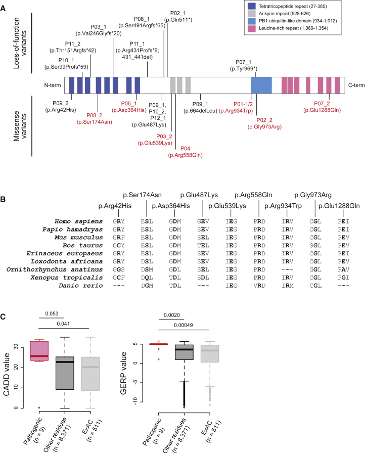
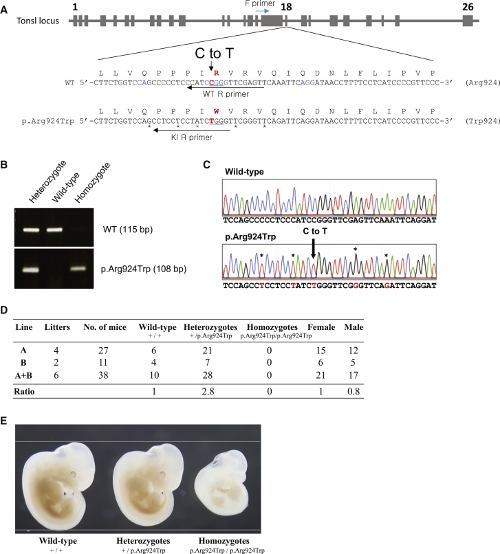
SPONASTRIME dysplasia is a rare, recessive skeletal dysplasia characterized by short stature, facial dysmorphism, and aberrant radiographic findings of the spine and long bone metaphysis. No causative genetic alterations for SPONASTRIME dysplasia have yet been determined. Using whole-exome sequencing (WES), we identified bi-allelic TONSL mutations in 10 of 13 individuals with SPONASTRIME dysplasia. TONSL is a multi-domain scaffold protein that interacts with DNA replication and repair factors and which plays critical roles in resistance to replication stress and the maintenance of genome integrity. We show here that cellular defects in dermal fibroblasts from affected individuals are complemented by the expression of wild-type TONSL. In addition, in vitro cell-based assays and in silico analyses of TONSL structure support the pathogenicity of those TONSL variants. Intriguingly, a knock-in (KI) Tonsl mouse model leads to embryonic lethality, implying the physiological importance of TONSL. Overall, these findings indicate that genetic variants resulting in reduced function of TONSL cause SPONASTRIME dysplasia and highlight the importance of TONSL in embryonic development and postnatal growth.

Keywords: DNA repair; DNA replication; SPONASTRIME dysplasia; TONSL; rare genetic diseases; short stature; skeletal dysplasia; whole-exome sequencing.


Figures

Figure 1
Figure 1
Characteristic Facial Appearance and Radiographic Findings of Individuals with SPONASTSRIME Dysplasia (A) Facial photos of individuals from three different ethnicities. They share midfacial hypoplasia, a depressed nasal root, a short and upturned nose, prognathism, and a relatively large head size with a prominent forehead. Individual P05 is shown at age 5 years, individual P06 at age 5 years, and individual P08 at age 13 years. (B) A lateral spine view of individual P02 at age 3 years and 8 months shows a taller anterior vertebral body and convex anterior endplates. A lateral spine view of individual P01-2 at age 13 years shows biconcavity of the endplates. Hip and knee anteroposterior views of individual P02 show metaphyseal irregularity and vertical striation and small, dysplastic epiphyses. Hip and knee anteroposterior views of individual P01-2 show residual avascular necrosis of the left femoral head and mixed dense striations and lucent area at the metaphysis of the knee.
Figure 2
Figure 2
TONSL Variants Identified in Individuals with SPONASTRIME Dysplasia (A) Pathogenic variants in TONSL found in SPONASTRIME-affected individuals are displayed on the TONSL protein, whose known functional domains are indicated. Variants in red were analyzed for variant functionality. (B) Evolutionary conservation of the nine missense variants found in SPONASTRIME-affected individuals. (C) Because they are parameters of an individual’s missense-variant (“Pathogenic”) functionality, CADD and GERP values were plotted, along with residues that overlapped with the ExAC Database (“ExAC”) and residues that were not polymorphic (“Other residues”).
Figure 3
Figure 3
Complementation of Dermal Fibroblasts from Individuals P03 and P04 through TONSL cDNA Transduction (A) TONSL protein levels and corresponding siRNA-mediated TONSL depletion of cells from individuals with SPONASTRIME dysplasia. BJ cells were used as a normal control. The basal TONSL level in cells derived from the affected individuals was lower than that in BJ cells, and all of the cells were successfully depleted by siRNA treatment. C = siControl and T = siTONSL. (B) Stable protein level of wild-type (WT) TONSL delivered through lentiviral transduction into fibroblasts derived from both individuals P03 and P04. EV and WT denote the empty vector and wild-type, respectively. The asterisk indicates the cross-reacting band. (C) Cells from affected individuals were sensitive to CPT but were rescued to a level comparable to that of BJ cells by the transduction of WT TONSL. The indicated cells, in triplicate, were exposed to different concentrations of CPT ranging from 0–32 nM. After 5 days, the cells were counted with a coulter counter, and the total number of cells at each concentration was divided by the number of untreated cells. FANCP/EV, a Fanconi anemia-derived cell line lacking Slx4, and FANCP/WT-SLX4, a genetically isogenic line of SLX4-complemented cells, were used as controls. The error bars represent the standard deviation (SD) of three replicates. (D) Representative images of CPT-induced DNA damage in Rad51 and γH2A.X foci. P03/EV, P03/WT-TONSL, P04/EV, and P04/WT-TONSL cells were treated with CPT (50 nM) overnight, then fixed and stained with anti-RAD51 and anti-γH2A.X antibodies. Impaired DNA damage induced RAD51 foci formation in those cells from affected individuals that were recovered to the level of normal cells via the transduction of WT-TONSL. (E) Statistical analysis of Rad51 foci. The percentage of Rad51 foci was calculated by taking the number of nuclei with n ≥ 10 Rad51 foci divided by the total number of nuclei from non-treated cells, or, for CPT-treated cells, divided by the number of γH2A.X-positive nuclei. The error bars represent the SD. (F) BrdU incorporation into DNA was reduced in P03/EV and P04/EV cells, which were rescued to a normal level by the transduction of WT-TONSL. The BrdU incorporation ratio was calculated by dividing the number of BrdU-incorporated cells by the number of total cells counted. The error bars represent the SD of three replicates. (G) DNA fiber analysis of CPT-treated cell lines derived from SPONASTRIME-affected individuals. BJ cells were used as a control. A schematic representation of the experiment is shown on the top, and the representative DNA fiber is shown on bottom. The red line indicates median value; ∗∗ p = 0.003 and ∗∗∗ p < 0.001.
Figure 4
Figure 4
In Vitro Cell-Based Assay of TONSL Variants Shows Defects in Cell Proliferation and Enhanced Sensitivity to Camptothecin (A) The protein levels of recombinant WT and TONSL variants from affected individuals transduced and stably expressed in HeLa cells upon endogenous TONSL depletion via siRNA that targeted the TONSL 3′ UTR. The TONSL antibody is able to detect both endogenous and HA-tagged TONSL, whereas the HA antibody is not able to detect endogenous TONSL. The asterisk indicates the cross-reacting band. (B) Cell-proliferation assay showing the varied proliferation rates of cells expressing mutant TONSL. EV cells were treated with either non-targeting siCtrl or siTONSL targeting the 3′ UTR. The rest of the cells were treated with siTONSL targeting the 3′ UTR. The cell proliferation rate was normalized to the cell number on day 1. The error bar represents the SD of three replicates. siCtrl = siControl. (C) TONSL variants in the absence of endogenous TONSL lead to checkpoint activation. Whole-cell extracts of HeLa cells expressing mutant TONSL were treated with siRNA targeting the 3′ UTR and analyzed by immunoblot with DNA-damage-response factors, phosphorylated CHK1 (pCHK1), or phosphorylated CHK2 (pCHK2). (D) TONSL variant cell lines were treated with 3′ UTR-targeting siRNA as in (B), then treated with increasing concentrations of CPT. The cells were stained with Hoechst, and the nuclei were counted 5 days after CPT treatment. Cell survival was normalized to that in vehicle-treated cells. The error bars represent the SD from three replicates.
Figure 5
Figure 5
Generation and Analysis of Homozygous Tonsl p.Arg924Trp Knock-In Mice via CRISPR-Cas9 (A) A schematic diagram showing the mouse Tonsl locus and the enlarged sequences of exon 18 of tTonsl, along with the sequences of the TonslArg924Trp allele. Blue letters in the TonslWT allele indicate a proto-spacer adjacent motif (PAM) sequence. Red letters indicate the substitution (C to T) target nucleotide in the TonslWT and the TonslArg924Trp alleles. The amino acid sequences from the TonslWT and the TonslArg924Trp alleles are shown at the top of the nucleotide sequences. The substituted nucleotides for both synonymous and targeted mutations are shown at the bottom of the TonslArg924Trp allele sequences by black and red asterisks (), respectively. Forward (F) and Reverse (R) PCR primers for genotyping are indicated. (B) Genotyping PCR for the TonslWT and the TonslArg924Trp alleles. The upper and bottom panels show the PCR products that were amplified from the TonslWT (115 bp) and the TonslArg924Trp (108 bp) alleles, respectively. (C) Chromatogram displaying the sequence of the TonslWT and the TonslArg924Trp loci. (D) Genotype distribution of offspring from heterozygous intercrosses. (E) Gross morphology of whole embryos at stage E11.5. Tonsl+/+ and Tonsl+/Arg924Trp mouse embryos show normal development, whereas a TonslArg924Trp/Arg924Trp embryo exhibits a growth retardation with the abnormal development of eyes and limbs.

References

    1. Fanconi S., Issler C., Giedion A., Prader A. The SPONASTRIME dysplasia: Familial short-limb dwarfism with saddle nose, spinal alterations and metaphyseal striation. Report of 4 siblings. Helv. Paediatr. Acta. 1983;38:267–280. - PubMed
    1. Lachman R.S., Stoss H., Spranger J. Sponastrime dysplasia. A radiologic-pathologic correlation. Pediatr. Radiol. 1989;19:417–424. - PubMed
    1. Langer L.O., Jr., Beals R.K., LaFranchi S., Scott C.I., Jr., Sockalosky J.J. Sponastrime dysplasia: Five new cases and review of nine previously published cases. Am. J. Med. Genet. 1996;63:20–27. - PubMed
    1. Masuno M., Nishimura G., Adachi M., Hotsubo T., Tachibana K., Makita Y., Imaizumi K., Kuroki Y. SPONASTRIME dysplasia: Report on a female patient with severe skeletal changes. Am. J. Med. Genet. 1996;66:429–432. - PubMed
    1. Langer L.O., Jr., Beals R.K., Scott C.I., Jr. Sponastrime dysplasia: Diagnostic criteria based on five new and six previously published cases. Pediatr. Radiol. 1997;27:409–414. - PubMed

Publication types

MeSH terms

Supplementary concepts