Review
. 2019 Mar 5;29(3):576-591.
doi: 10.1016/j.cmet.2019.01.015. Epub 2019 Feb 14.

Cancer Metabolism Drives a Stromal Regenerative Response

Affiliations
Review

Cancer Metabolism Drives a Stromal Regenerative Response

Simon Schwörer et al. Cell Metab. .

Abstract

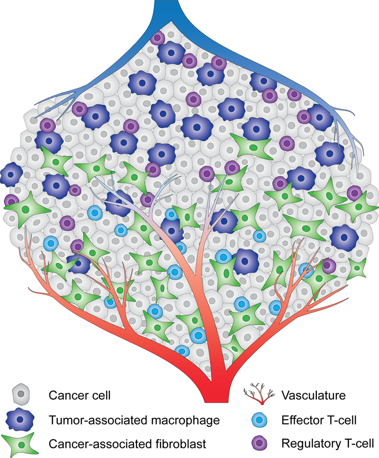
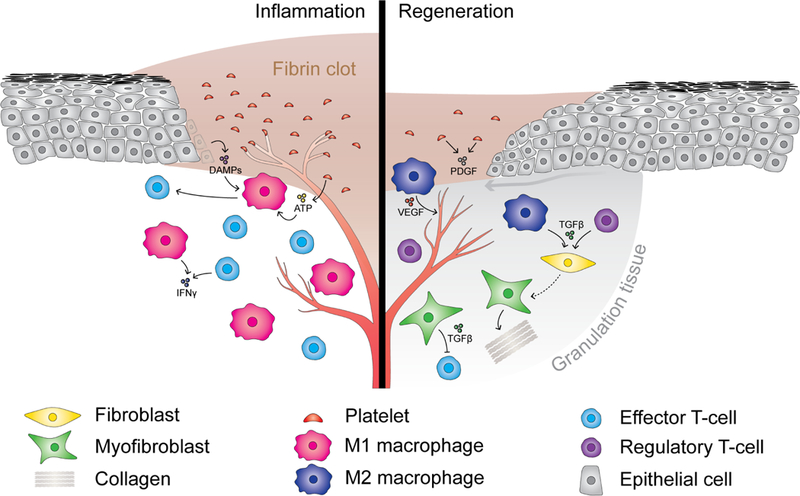
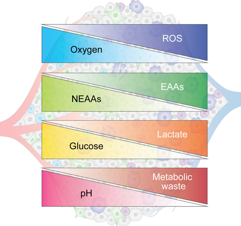
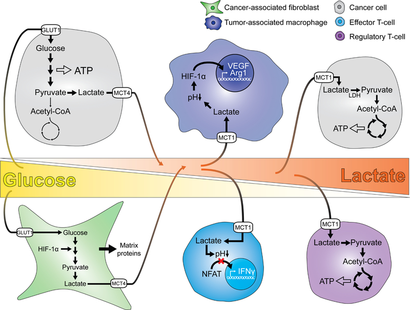
The metabolic reprogramming associated with malignant transformation has led to a growing appreciation of the nutrients required to support anabolic cell growth. Less well studied is how cancer cells satisfy those demands in vivo, where they are dispersed within a complex microenvironment. Tumor-associated stromal components can support tumor growth by providing nutrients that supplement those provided by the local vasculature. These non-malignant stromal cells are phenotypically similar to those that accumulate during wound healing. Owing to their immediate proximity, stromal cells are inevitably affected by the metabolic activity of their cancerous neighbors. Until recently, a role for tumor cell metabolism in influencing the cell fate decisions of neighboring stromal cells has been underappreciated. Here, we propose that metabolites consumed and released by tumor cells act as paracrine factors that regulate the non-malignant cellular composition of a developing tumor by driving stromal cells toward a regenerative response that supports tumor growth.

Keywords: cancer metabolism; cancer-associated fibroblasts; effector T cells; metabolism; regeneration; regulatory T cells; tumor microenvironment; tumor-associated macrophages; wound healing.


Conflict of interest statement

Conflict of interest

C.B.T. is a founder of Agios Pharmaceuticals and a member of its scientific advisory board. He is a former member of the Board of Directors and a stockholder of both Merck and Charles River Laboratories. He is a named inventor on patents related to cellular metabolism.

Figures

Figure 1
Figure 1. The tumor microenvironment.
Cancer cells are dispersed within a complex microenvironment. Multiple nonmalignant cell types are found in a bulk tumor, including fibroblasts, T-cells and macrophages. While cancer-associated fibroblasts and effector T-cells are preferentially found in the proximity of the vasculature, tumor-associated macrophages and regulatory T-cells accumulate in hypovascular tumor regions. Other non-malignant cell types are present in tumor stroma but not depicted here to aid visualization.
Figure 2
Figure 2. The wound healing response.
The inflammatory phase of the wound healing response (left) is characterized by accumulation of pro-inflammatory immune cells, including M1 macrophages and effector T-cells. Damage-associated molecular patterns (DAMPs) including ATP released from dying epithelial cells and platelets initially recruit monocytes to the site of tissue damage where they differentiate into pro-inflammatory M1 macrophages. M1 macrophages clear necrotic tissue and secrete cytokines that recruit effector T-cells. Following clearance of cell debris, M2 macrophages and regulatory T-cells accumulate (right), secreting cytokines and growth factors such as TGFβ that induce activation and differentiation of fibroblasts into myofibroblasts. Myofibroblasts contribute to the formation of granulation tissue and wound repair by secretion of ECM proteins such as collagens, and by secretion of immunosuppressive factors including TGFβ. Angiogenesis is stimulated by VEGF secreted by M2 macrophages.
Figure 3
Figure 3. Metabolic gradients in tumors.
The metabolic activity of cancer cells and stromal cells as well as proximity to the vasculature contribute to the formation of metabolic gradients within tumors. In hypovascularized tumor core regions (right), glucose, glutamine and other nonessential amino acids (NEAAs) are depleted, while certain essential amino acids (EAAs) and metabolic waste products such as lactate accumulate. These areas are also characterized by elevated levels of reactive oxygen species (ROS) and acidic pH.
Figure 4
Figure 4. Lactate’s effects on tumor and stromal cells.
Glucose is present in high levels in proximity to the vasculature (left) and is consumed by tumor cells and cancer-associated fibroblasts. Glucose consumption is coupled to lactate secretion, resulting in lactate accumulation with increasing distance from the efferent vasculature. In poorly vascularized regions (right), cancer cells and regulatory T-cells are able to metabolize lactate to sustain energy homeostasis. In contrast, lactate exerts paracrine effects on tumor-associated macrophages and effector T-cells, stimulating differentiation of macrophages into an immunosuppressive M2-like phenotype and suppressing T-cell effector function.

References

    1. Achreja A, Zhao H, Yang L, Yun TH, Marini J, and Nagrath D (2017). ExoMFA – A 13C metabolic flux analysis framework to dissect tumor microenvironmentsecreted exosome contributions towards cancer cell metabolism. Metab. Eng 43, 156–172. - PMC - PubMed
    1. Adler M, Mayo A, Zhou X, Franklin RA, Jacox JB, Medzhitov R, and Alon U (2018). Endocytosis as a stabilizing mechanism for tissue homeostasis. Proc. Natl. Acad. Sci 115, E1926–E1935. - PMC - PubMed
    1. Ammirante M, Shalapour S, Kang Y, Jamieson CAM, and Karin M (2014). Tissue injury and hypoxia promote malignant progression of prostate cancer by inducing CXCL13 expression in tumor myofibroblasts. Proc. Natl. Acad. Sci 111, 14776–14781. - PMC - PubMed
    1. Andrianifahanana M, Hernandez DM, Yin X, Kang JH, Jung MY, Wang Y, Yi ES, Roden AC, Limper AH, and Leof EB (2016). Profibrotic up-regulation of glucose transporter 1 by TGF-β involves activation of MEK and mammalian target of rapamycin complex 2 pathways. FASEB J 30, 3733–3744. - PMC - PubMed
    1. Angelin A, Gil-de-Gómez L, Dahiya S, Jiao J, Guo L, Levine MH, Wang Z, Quinn WJ, Kopinski PK, Wang L, et al. (2017). Foxp3 Reprograms T Cell Metabolism to Function in Low-Glucose, High-Lactate Environments. Cell Metab 25, 1282–1293.e7. - PMC - PubMed

Publication types

LinkOut - more resources