

[Effects of loupes and microscope on laminate veneer preparation]
- PMID: 30773552
- PMCID: PMC7433548
- DOI: 10.19723/j.issn.1671-167X.2019.01.018
[Effects of loupes and microscope on laminate veneer preparation]
Abstract
Objective: To assess and compare the effects of loupes and microscope on laminate veneer preparation of the first practitioner from the aspects of efficiency, quality and accuracy of preparation, and preference.
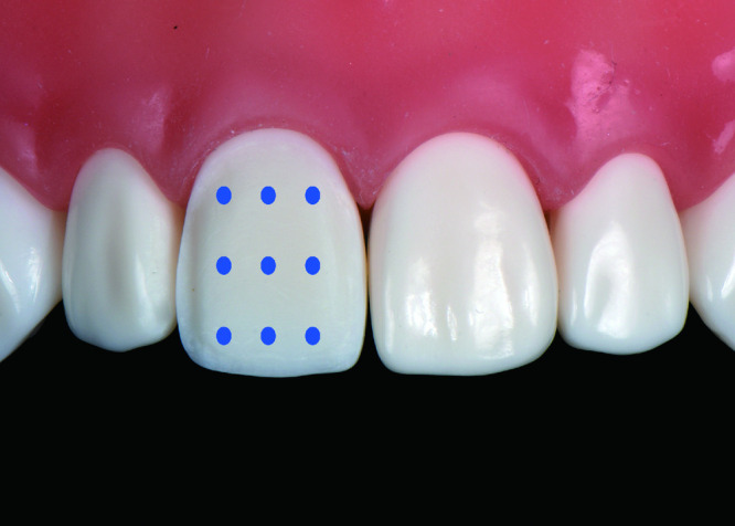
Methods: Twenty young prosthodontists from the Department of Prosthodontics, Peking University School and Hospital of Stomatology were recruited into this study, which was prospective, single blind, self-control trials. The participants had no experience of using dental magnification devices. They prepared laminate veneers in the artificial dental model, under routine visual field (control group), 2.5× headwear loupes (loupes group), and 8× operating microscope (microscopic group) by turning. The time for tooth preparation was recorded. Thereafter, subjective assessments of efficiency, quality of preparation and preference were performed by themselves using visual analogue score (VAS). Expert assessments of quality and accuracy of preparation were performed by two professors using stereomicroscope and digital technique respectively.
Results: In terms of efficiency, the subjective scores for the control group, loupes group and microscopic group were 7.15±1.73, 8.10±0.91 and 5.40±2.04, respectively. There was significant difference between the loupes group and microscopic group (P<0.05). The time of tooth preparation for the control group, loupes group and microscopic group was (430.10±163.04) s, (393.90±157.27) s and (441.95±164.18) s, respectively. There was significant difference between the loupes group and microscopic group (P<0.05). The loupes group was more efficient than the microscopic group. In terms of the quality of preparations, the subjective scores for the control group, loupes group and microscopic group were 6.55±2.09, 7.85±0.99 and 6.25±1.77, respectively. There was significant difference between the loupes group and microscopic group (P<0.05). The expert evaluations for the control group, loupes group and microscopic group were 12.20±1.67, 12.50±1.70 and 11.35±2.60, respectively. There was significant difference between the loupes group and microscopic group (P<0.05). The loupes group had higher quality than the microscopic group. In terms of the accuracy of preparations, the control group, loupes group and microscopic group of incisal 1/3 were (0.107±0.097) mm, (0.142±0.118) mm and (0.123±0.087) mm, respectively, of middle 1/3 were (0.128±0.073) mm, (0.113±0.105) mm and (0.125±0.077) mm, respectively, and of cervical 1/3 were (0.075±0.054) mm, (0.068±0.044) mm and (0.058±0.047) mm, respectively. There was no significant difference among the three groups (P>0.05). In terms of the preference, the subjective scores for the control group, loupes group and microscopic group were 6.55±2.31, 8.60±1.10 and 5.80±2.07, respectively. There was significant difference between the loupes group and microscopic group (P<0.05). The participants had the highest preference for loupes.
Conclusion: For the first practitioners, loupes is better than microscope for laminate veneer preparation.
目的: 研究和比较初学者应用放大镜与应用显微镜进行瓷贴面牙体预备的效果,从操作效率、预备体质量、预备准确度以及喜好度等方面比较放大镜和显微镜的应用价值。
方法: 从北京大学口腔医院修复科选择20名口腔修复医生进行前瞻性、单盲、自身对照试验,试验对象无使用放大镜或显微镜的经验。每人依次在常规视野下(空白对照组)、2.5倍头戴式放大镜下(放大镜组)和8倍医用显微镜下(显微镜组)在仿头模内完成右上中切牙开窗型瓷贴面牙体预备,试验过程中记录牙体预备所需的时间。操作完成后,由医生本人利用视觉模拟评分法(vi-sual analogue score,VAS)对操作效率、预备体质量和喜好度进行主观评分,由第三方专家在体视显微镜下对瓷贴面预备体的质量进行评分,并利用数字化方法对预备准确性进行评价。
结果: 操作效率方面,对照组、放大镜组和显微镜组的主观VAS评分分别为7.15±1.73、8.10±0.91、5.40±2.04,放大镜组与显微镜组间的差异有统计学意义(P<0.05);客观操作时间三组分别为(430.10±163.04) s、(393.90±157.27) s、(441.95±164.18) s,放大镜组与显微镜组间的差异有统计学意义(P<0.05);放大镜组比显微镜组操作效率高。预备体质量方面,对照组、放大镜组和显微镜组的主观VAS评分分别为6.55±2.09、7.85±0.99、6.25±1.77,放大镜组与显微镜组间的差异有统计学意义(P<0.05);专家评分分别为12.20±1.67、12.50±1.70、11.35±2.60,放大镜组与显微镜组间的差异有统计学意义(P<0.05);放大镜组的预备体质量优于显微镜组。预备准确度方面,对照组、放大镜组和显微镜组的唇面切1/3分别为(0.107±0.097) mm、(0.142±0.118) mm、(0.123±0.087) mm,唇面中1/3分别为(0.128±0.073) mm、(0.113±0.105) mm、(0.125±0.077) mm,唇面颈1/3分别为(0.075±0.054) mm、(0.068±0.044) mm、(0.058±0.047) mm,三组间每个区域的差异均无统计学意义(P>0.05)。喜好度方面,对照组、放大镜组和显微镜组的主观VAS评分分别为6.55±2.31、8.60±1.10、5.80±2.07,放大镜组与显微镜组间的差异有统计学意义(P<0.05),放大镜组最受欢迎。
结论: 针对初学者而言,放大镜比显微镜用于瓷贴面牙体预备的效果更好。
Conflict of interest statement
The authors have declared that no competing interests exist.
作者已声明无竞争性利益关系。
Figures
Similar articles
-
[Effects of loupes and microscope on the prosthodontist's posture from ergonomic aspects].Beijing Da Xue Xue Bao Yi Xue Ban. 2020 Oct 18;52(5):948-951. doi: 10.19723/j.issn.1671-167X.2020.05.026. Beijing Da Xue Xue Bao Yi Xue Ban. 2020. PMID: 33047735 Free PMC article. Chinese.
-
Effects of loupes and microscopes on a dental technician's working posture from ergonomic aspects.Hua Xi Kou Qiang Yi Xue Za Zhi. 2022 Jul 25;40(4):428-435. doi: 10.7518/hxkq.2022.04.009. Hua Xi Kou Qiang Yi Xue Za Zhi. 2022. PMID: 38596959 Free PMC article. Chinese, English.
-
In vitro evaluation of accuracy and precision of automated robotic tooth preparation system for porcelain laminate veneers.J Prosthet Dent. 2015 Aug;114(2):229-35. doi: 10.1016/j.prosdent.2015.02.021. Epub 2015 May 5. J Prosthet Dent. 2015. PMID: 25957239
-
Survival rates for porcelain laminate veneers with special reference to the effect of preparation in dentin: a literature review.J Esthet Restor Dent. 2012 Aug;24(4):257-65. doi: 10.1111/j.1708-8240.2012.00517.x. Epub 2012 May 29. J Esthet Restor Dent. 2012. PMID: 22863131 Review.
-
Incisal preparation design for ceramic veneers: A critical review.J Am Dent Assoc. 2018 Jan;149(1):25-37. doi: 10.1016/j.adaj.2017.08.031. J Am Dent Assoc. 2018. PMID: 29304908 Review.
Cited by
-
Do magnification loupes affect the precision of cavity preparations made by undergraduates? A randomized crossover study.BMC Oral Health. 2022 May 19;22(1):189. doi: 10.1186/s12903-022-02232-z. BMC Oral Health. 2022. PMID: 35590298 Free PMC article. Clinical Trial.
-
Impact of using magnifying dental loupes on clinical performance during tooth preparation: A systematic review.J Clin Exp Dent. 2024 Feb 1;16(2):e186-e197. doi: 10.4317/jced.61098. eCollection 2024 Feb. J Clin Exp Dent. 2024. PMID: 38496818 Free PMC article. Review.
-
Application of medical magnifying loupes in diagnosis of oral mucosal diseases.Zhejiang Da Xue Xue Bao Yi Xue Ban. 2021 Apr 25;50(2):205-211. doi: 10.3724/zdxbyxb-2021-0132. Zhejiang Da Xue Xue Bao Yi Xue Ban. 2021. PMID: 34137223 Free PMC article. English.
-
Effects of magnification on restorative dental preparation performance: a scoping review and level of evidence mapping.Clin Oral Investig. 2024 Jul 25;28(8):447. doi: 10.1007/s00784-024-05852-7. Clin Oral Investig. 2024. PMID: 39052037
-
[Clinical pathway and preparation method of high-precision tooth shoulder platform].Hua Xi Kou Qiang Yi Xue Za Zhi. 2020 Dec 1;38(6):712-717. doi: 10.7518/hxkq.2020.06.019. Hua Xi Kou Qiang Yi Xue Za Zhi. 2020. PMID: 33377353 Free PMC article. Chinese.
References
-
- Carr GB, Murgel CA. The use of the operating microscope in endodontics. Dent Clin North Am. 2010;54(2):191–214. - PubMed
-
- Eichenberger M, Perrin P, Ramseyer ST, et al. Visual acuity and experience with magnification devices in Swiss dental practices. Oper Dent. 2015;40(4):E142–E149. - PubMed
-
- Perrin P, Eichenberger M, Neuhaus KW, et al. A visual acuity and magnification devices in dentistry. Swiss Dent J. 2016;126(3):222–235. - PubMed
-
- Sitbon Y, Attathom T. Minimal intervention dentistry II: Part 6. Microscope and microsurgical techniques in periodontics. British Dent J. 2014;216(9):503–509. - PubMed
-
- Sitbon Y, Attathom T, St-Georges AJ. Minimal intervention dentistry II: part 1. Contribution of the operating microscope to dentistry. British Dent J. 2014;216(3):125–130. - PubMed