. 2019 Jan 30:(143):10.3791/58715.
doi: 10.3791/58715.

Determining 3'-Termini and Sequences of Nascent Single-Stranded Viral DNA Molecules during HIV-1 Reverse Transcription in Infected Cells

Affiliations

Determining 3'-Termini and Sequences of Nascent Single-Stranded Viral DNA Molecules during HIV-1 Reverse Transcription in Infected Cells

Darja Pollpeter et al. J Vis Exp. .

Abstract

Monitoring of nucleic acid intermediates during virus replication provides insights into the effects and mechanisms of action of antiviral compounds and host cell proteins on viral DNA synthesis. Here we address the lack of a cell-based, high-coverage, and high-resolution assay that is capable of defining retroviral reverse transcription intermediates within the physiological context of virus infection. The described method captures the 3'-termini of nascent complementary DNA (cDNA) molecules within HIV-1 infected cells at single nucleotide resolution. The protocol involves harvesting of whole cell DNA, targeted enrichment of viral DNA via hybrid capture, adaptor ligation, size fractionation by gel purification, PCR amplification, deep sequencing, and data analysis. A key step is the efficient and unbiased ligation of adaptor molecules to open 3'-DNA termini. Application of the described method determines the abundance of reverse transcripts of each particular length in a given sample. It also provides information about the (internal) sequence variation in reverse transcripts and thereby any potential mutations. In general, the assay is suitable for any questions relating to DNA 3'-extension, provided that the template sequence is known.


Figures

Figure 1
Figure 1. Diagram showing the first steps of HIV-1 reverse transcription.
The process starts with annealing of tRNA(Lys,3) (orange) to the primer binding site (PBS) in the genomic viral RNA (step 1), which allows the initiation and elongation of viral cDNA (blue, step 2). Concomitantly, the template genomic RNA is degraded by RNaseH activity of RT (step 3). The first full intermediate in the process of reverse transcription is the minus-strand strong-stop (-)sss cDNA, which is complete when the RT catalyzed polymerization reaches the 5'-terminus of the gRNA repeat (R) region (step 3). The (-)sss intermediate is transferred to the 3'-terminus of the genomic RNA template by annealing to the complementary 3'-long terminal repeat (LTR) R region. From here, polymerization continues (step 4). In the described method the reverse transcription progression is determined by mapping the exact length of the nascent viral cDNA (blue). PPT, polypurine tract; U5, unique 5'-sequence; U3, unique 3'-sequence. This figure is republished from a previous publication. Please click here to view a larger version of this figure.
Figure 2
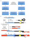
Figure 2. Workflow outline and schematics of the adapter ligation and PCR amplification strategy.
(a) Workflow outlining main steps of the described technique to determine 3'-termini of HIV-1 reverse transcripts in infected cells. The figure is adapted from a previous publication. (b) Schematic of the adaptor ligation and PCR amplification strategy. Nascent cDNA molecules of varying length that have been purified in previous steps are ligated to a single-stranded DNA adaptor using T4 DNA ligase. The hairpin adaptor (named "full Kwok + MiSeq", see Table 1 design was inspired by Kwok et al.. The adaptor carries a random 6 nt barcode sequence, which allows for base-pairing to facilitate ligation and simultaneously serves as an identifier for unique reads. The 3'-termini of the adaptor carries a spacer (SpC3) to prevent self-ligation. Ligated products are separated from excess adaptor by denaturing polyacrylamide gel electrophoresis (PAGE). Nucleic acids in the gel are stained and cut into three separate, equal-size gel pieces in the area from above the adaptor to the well as done in. After elution, precipitation, and resuspension, the products are PCR amplified with primers annealing to the known sequence of the adaptor (primer 1, multiplex oligonucleotide kit, see Table of Materials) and a primer carrying the first 22 nt of the HIV-1 5'-LTR sequence immediately following the tRNA (primer 2, MP1.0 + 22HIV). The 5'-termini of the chosen primers carry adaptors for the chosen sequencing platform (P5 and P7) as well as an index sequence to distinguish individual samples run in the same library. Starting points of the sequencing read primers are indicated. The blue box indicates the region of interest to determine the original 3'-termini of the captured molecule. This figure is adapted from a previous publication. Please click here to view a larger version of this figure.
Figure 3
Figure 3. Representative results.
(a) The total read count of representative samples processed with the described protocol. This includes all sequences that were identified as unique reads of HIV-1 molecules with their 3'-termini within the first 635 nt of the minus strand cDNA (up to the PPT, see Figure 1). Infection with HIV-1 not carrying A3G yields the highest number of reads, whereas A3G inhibits cDNA synthesis and thereby reduces the total read count. Uninfected cells served as a negative control, while a set of synthetic oligonucleotides provides a positive control. b) The relative abundance of cDNAs for each length between nt positions 23 and 182 (full-length -sss cDNA is 180 to 182 nt) of the HIV-1NL4.3 sequence (x-axis) is shown in blue histograms (scale on the left y-axis). The relative abundance of cDNA was calculated from the absolute number of sequences terminating at a given nucleotide within the -sss cDNA sequence divided by the sum of all reads measuring 182nt or less. Shown in dashed red lines are the percentages of reads carrying C-to-T/U mutations at the respective position (scale on the right y-axes). Figure 3b is republished from a previous publication. Please click here to view a larger version of this figure.
Figure 4
Figure 4. Representative results of control samples.
Shown are two profiles for pools containing equimolar amounts of 17 different length synthetic oligonucleotides. These oligonucleotides have sequences from HIV-1NL4.3 and were selected to cover various lengths and present all 4 bases as a 3'-nucleotide (see Table 2). The top graph shows the positive control sample from Figure 3a. No significant bias towards molecule length or the open 3'-termini is detected. The bottom graph shows a different library run, which produced a minor length bias in sequencing. In this case, it is advisable to apply a normalization factor, which is derived from the slope (shown in pink) that represents the size bias. This figure is republished from a previous publication. Please click here to view a larger version of this figure.

References

    1. Herschhorn A, Hizi A. Retroviral reverse transcriptases. Cellular and Molecular Life Sciences. 2010;67(16):2717–2747. - PMC - PubMed
    1. Hu WS, Hughes SH. HIV-1 reverse transcription. Cold Spring Harbor Perspectives in Medicine. 2012;2(10) - PMC - PubMed
    1. Levin JG, Mitra M, Mascarenhas A, Musier-Forsyth K. Role of HIV-1 nucleocapsid protein in HIV-1 reverse transcription. RNA Biology. 2010;7(6):754–774. - PMC - PubMed
    1. Menendez-Arias L, Sebastian-Martin A, Alvarez M. Viral reverse transcriptases. Virus Research. 2016 - PubMed
    1. Telesnitsky A, Goff SP. In: Retroviruses. Coffin JM, Hughes SH, Varmus HE, editors. 1997. - PubMed

Publication types