

Treatment of large avascular retinal pigment epithelium detachments in age-related macular degeneration with aflibercept, photodynamic therapy, and triamcinolone acetonide
- PMID: 30774304
- PMCID: PMC6362940
- DOI: 10.2147/OPTH.S188315
Treatment of large avascular retinal pigment epithelium detachments in age-related macular degeneration with aflibercept, photodynamic therapy, and triamcinolone acetonide
Abstract
Purpose: To evaluate the use of aflibercept, triamcinolone acetonide, and photodynamic therapy (PDT) in the treatment of avascular pigment epithelium detachments (aPEDs).
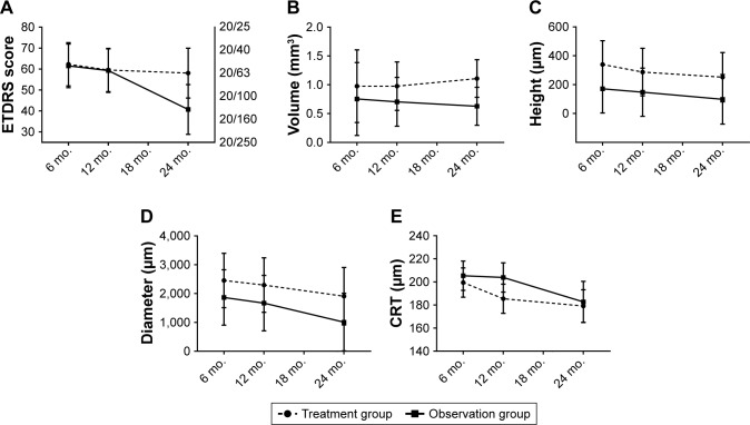
Patients and methods: Patients with treatment-naïve aPEDs ≥1,500 µm in diameter were randomized to treatment or observation. Treatment consisted of 6 monthly intravitreal injections of aflibercept. If the aPED persisted, the patients were treated with half-fluence PDT in combination with intravitreal triamcinolone acetonide and aflibercept. The primary outcome was change of best-corrected visual acuity (BCVA) after 24 months of follow-up. Secondary outcomes were changes in pigment epithelium volume, height and diameter, central retinal thickness, and number of patients developing choroidal neovascularization or geographic atrophy (GA).
Results: Treatment and inclusion of patients were stopped after an interim analysis of 6-month data because 75% of the aPEDs were in different stages of GA. Nine patients with aPED were included in the study, of these one patient was excluded because of bilateral central serous chorioretinopathy. The remaining eight had drusenoid aPEDs. After 24 months of follow-up, estimated means of BCVA decreased by 4.2 and 20.8 letters in the treatment and observation group, respectively. This decrease over time was not significantly different between groups (P=0.140, 95% CI -5.3, 38.6). Estimated means of PED volume, height, diameter, and central retinal thickness were not significantly different between groups. Choroidal neovascularization and retinal pigment epithelium tear developed in one patient in the treatment group. One patient in the treatment group and two patients in the observation group progressed to complete retinal pigment epithelium and outer retinal atrophy. A decrease in PED volume was associated with the development of complete retinal pigment epithelium and outer retinal atrophy (P=0.029).
Conclusion: This small trial indicates that multitargeted, primarily antiangiogenic therapy does not favorably alter the natural course of drusenoid aPEDs.
Keywords: PDT; anti-VEGF; drusenoid; geographic atrophy.
Conflict of interest statement
Disclosure The authors report no conflicts of interest in this work.
Figures
Similar articles
-
Prospective PED-study of intravitreal aflibercept for refractory vascularized pigment epithelium detachment due to age-related macular degeneration: morphologic characteristics of non-responders in optical coherence tomography.Graefes Arch Clin Exp Ophthalmol. 2020 Jul;258(7):1411-1417. doi: 10.1007/s00417-020-04675-y. Epub 2020 Apr 18. Graefes Arch Clin Exp Ophthalmol. 2020. PMID: 32306096 Free PMC article.
-
Response of vascular pigment epithelium detachment due to age-related macular degeneration to monthly treatment with ranibizumab: the prospective, multicentre RECOVER study.Acta Ophthalmol. 2017 Nov;95(7):683-689. doi: 10.1111/aos.13359. Epub 2017 Jan 13. Acta Ophthalmol. 2017. PMID: 28084038 Clinical Trial.
-
Vascularized retinal pigment epithelial detachment in age-related macular degeneration: treatment and RPE tear incidence.Graefes Arch Clin Exp Ophthalmol. 2012 Sep;250(9):1283-92. doi: 10.1007/s00417-012-1955-2. Epub 2012 Feb 21. Graefes Arch Clin Exp Ophthalmol. 2012. PMID: 22350060
-
Clinical evidence of intravitreal triamcinolone acetonide in the management of age-related macular degeneration.Curr Drug Targets. 2011 Feb;12(2):149-72. doi: 10.2174/138945011794182746. Curr Drug Targets. 2011. PMID: 20887246 Review.
-
Photodynamic therapy for polypoidal choroidal vasculopathy.Prog Retin Eye Res. 2013 Nov;37:182-99. doi: 10.1016/j.preteyeres.2013.09.003. Epub 2013 Oct 15. Prog Retin Eye Res. 2013. PMID: 24140257 Review.
Cited by
-
Retinal Pigment Epithelial Detachment in Age-Related Macular Degeneration.Ophthalmol Ther. 2020 Dec;9(4):739-756. doi: 10.1007/s40123-020-00291-5. Epub 2020 Aug 18. Ophthalmol Ther. 2020. PMID: 32809132 Free PMC article. Review.
-
Long-term outcomes of drusenoid retinal pigment epithelium detachment in eyes with age-related macular degeneration.Indian J Ophthalmol. 2025 Jun 1;73(6):843-846. doi: 10.4103/IJO.IJO_1966_24. Epub 2025 May 28. Indian J Ophthalmol. 2025. PMID: 40434460 Free PMC article.
References
-
- Klein BE, Klein R. Cataracts and macular degeneration in older Americans. Arch Ophthalmol. 1982;100(4):571–573. - PubMed
-
- Bourne RR, Jonas JB, Flaxman SR, et al. Prevalence and causes of vision loss in high-income countries and in eastern and central Europe: 1990–2010. Br J Ophthalmol. 2014;98(5):629–638. - PubMed
-
- Schmidt-Erfurth U, Waldstein SM. A paradigm shift in imaging biomarkers in neovascular age-related macular degeneration. Prog Retin Eye Res. 2016;50:1–24. - PubMed
-
- Zayit-Soudry S, Moroz I, Loewenstein A. Retinal pigment epithelial detachment. Surv Ophthalmol. 2007;52(3):227–243. - PubMed
LinkOut - more resources
Full Text Sources
