

Bisperoxovanadium protects against spinal cord injury by regulating autophagy via activation of ERK1/2 signaling
- PMID: 30774313
- PMCID: PMC6362923
- DOI: 10.2147/DDDT.S187878
Bisperoxovanadium protects against spinal cord injury by regulating autophagy via activation of ERK1/2 signaling
Abstract
Background: Spinal cord injury (SCI) is a disease of the central nervous system with few restorative treatments. Autophagy has been regarded as a promising therapeutic target for SCI. The inhibitor of phosphatase and tensin homolog deleted on chromosome ten (PTEN) bisperoxovanadium (bpV[pic]) had been claimed to provide a neuroprotective effect on SCI; but the underlying mechanism is still not fully understood.
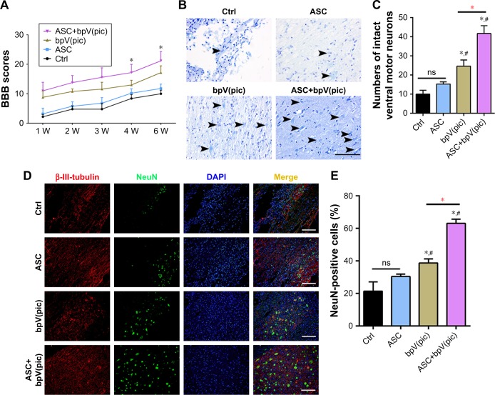
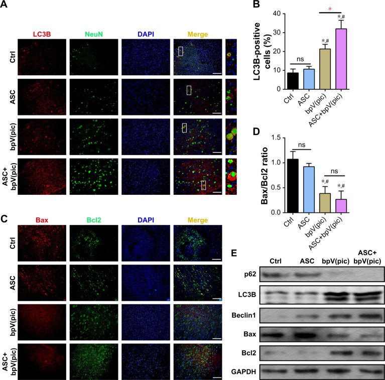
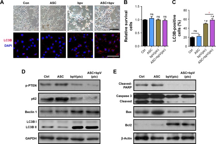
Materials and methods: Acute SCI model were generated with SD Rats and were treated with control, acellular spinal cord scaffolds (ASC) obtained from normal rats, bpV(pic), and combined material of ASC and bpV(pic). We used BBB score to assess the motor function of the rats and the motor neurons were stained with Nissl staining. The expressions of the main autophagy markers LC3B, Beclin1 and P62, expressions of apoptosis makers Bax, Bcl2, PARP and Caspase 3 were detected with IF or Western Blot analysis.
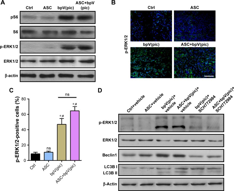
Results: The bpV(pic) showed significant improvement in functional recovery by activating autophagy and accompanied by decreased neuronal apoptosis; combined ASC with bpV(pic) enhanced these effects. In addition, after treatment with ERK1/2 inhibitor SCH772984, we revealed that bpV(pic) promotes autophagy and inhibits apoptosis through activating ERK1/2 signaling after SCI.
Conclusion: These results illustrated that the bpV(pic) protects against SCI by regulating autophagy via activation of ERK1/2 signaling.
Keywords: ERK1/2 signaling; apoptosis; autophagy; bisperoxovanadium; spinal cord injury.
Conflict of interest statement
Disclosure The authors report no conflicts of interest in this work.
Figures
Similar articles
-
Bisperoxovanadium Mediates Neuronal Protection through Inhibition of PTEN and Activation of PI3K/AKT-mTOR Signaling after Traumatic Spinal Injuries.J Neurotrauma. 2019 Sep 15;36(18):2676-2687. doi: 10.1089/neu.2018.6294. Epub 2019 Mar 28. J Neurotrauma. 2019. PMID: 30672370 Free PMC article.
-
PTEN inhibitor bisperoxovanadium protects oligodendrocytes and myelin and prevents neuronal atrophy in adult rats following cervical hemicontusive spinal cord injury.Neurosci Lett. 2014 Jun 24;573:64-8. doi: 10.1016/j.neulet.2014.02.039. Epub 2014 Feb 26. Neurosci Lett. 2014. PMID: 24582904 Free PMC article.
-
Bisperoxovanadium induces M2-type macrophages and promotes functional recovery after spinal cord injury.Mol Immunol. 2019 Dec;116:56-62. doi: 10.1016/j.molimm.2019.09.022. Epub 2019 Oct 9. Mol Immunol. 2019. PMID: 31605961
-
[Progress of researches on acupuncture treatment of spinal cord injury by regulating programmed cell death].Zhen Ci Yan Jiu. 2024 Dec 25;49(12):1325-1332. doi: 10.13702/j.1000-0607.20240310. Zhen Ci Yan Jiu. 2024. PMID: 39681492 Review. Chinese.
-
Modulation of autophagy for neuroprotection and functional recovery in traumatic spinal cord injury.Neural Regen Res. 2020 Sep;15(9):1601-1612. doi: 10.4103/1673-5374.276322. Neural Regen Res. 2020. PMID: 32209759 Free PMC article. Review.
Cited by
-
Valproic Acid: A Potential Therapeutic for Spinal Cord Injury.Cell Mol Neurobiol. 2021 Oct;41(7):1441-1452. doi: 10.1007/s10571-020-00929-9. Epub 2020 Jul 28. Cell Mol Neurobiol. 2021. PMID: 32725456 Free PMC article. Review.
-
Cryogenic 3D Printing of ß-TCP/PLGA Composite Scaffolds Incorporated With BpV (Pic) for Treating Early Avascular Necrosis of Femoral Head.Front Bioeng Biotechnol. 2022 Jan 18;9:748151. doi: 10.3389/fbioe.2021.748151. eCollection 2021. Front Bioeng Biotechnol. 2022. PMID: 35118053 Free PMC article.
-
Ezetimibe protects against spinal cord injury by regulating autophagy and apoptosis through inactivation of PI3K/AKT/mTOR signaling.Am J Transl Res. 2020 Jun 15;12(6):2685-2694. eCollection 2020. Am J Transl Res. 2020. PMID: 32655800 Free PMC article.
-
Pyroptosis, ferroptosis, and autophagy in spinal cord injury: regulatory mechanisms and therapeutic targets.Neural Regen Res. 2025 Oct 1;20(10):2787-2806. doi: 10.4103/NRR.NRR-D-24-00112. Epub 2024 Jul 29. Neural Regen Res. 2025. PMID: 39101602 Free PMC article.
-
Crosstalk between exosomes and autophagy in spinal cord injury: fresh positive target for therapeutic application.Cell Tissue Res. 2023 Jan;391(1):1-17. doi: 10.1007/s00441-022-03699-6. Epub 2022 Nov 16. Cell Tissue Res. 2023. PMID: 36380098 Free PMC article. Review.
References
-
- McDonald JW, Sadowsky C. Spinal-cord injury. Lancet. 2002;359(9304):417–425. - PubMed
-
- Young W. Spinal cord regeneration. Cell Transplant. 2014;23(4–5):573–611. - PubMed
-
- Carlson GD, Gorden C. Current developments in spinal cord injury research. Spine J. 2002;2(2):116–128. - PubMed
-
- Thuret S, Moon LD, Gage FH. Therapeutic interventions after spinal cord injury. Nat Rev Neurosci. 2006;7(8):628–643. - PubMed
-
- Norenberg MD, Smith J, Marcillo A. The pathology of human spinal cord injury: defining the problems. J Neurotrauma. 2004;21(4):429–440. - PubMed
MeSH terms
Substances
LinkOut - more resources
Full Text Sources
Medical
Research Materials
Miscellaneous
