

microRNA-628 inhibits the proliferation of acute myeloid leukemia cells by directly targeting IGF-1R
- PMID: 30774377
- PMCID: PMC6357892
- DOI: 10.2147/OTT.S192137
microRNA-628 inhibits the proliferation of acute myeloid leukemia cells by directly targeting IGF-1R
Retraction in
-
microRNA-628 Inhibits the Proliferation of Acute Myeloid Leukemia Cells by Directly Targeting IGF-1R [Retraction].Onco Targets Ther. 2021 Sep 19;14:4847-4848. doi: 10.2147/OTT.S339844. eCollection 2021. Onco Targets Ther. 2021. PMID: 34584423 Free PMC article.
Abstract
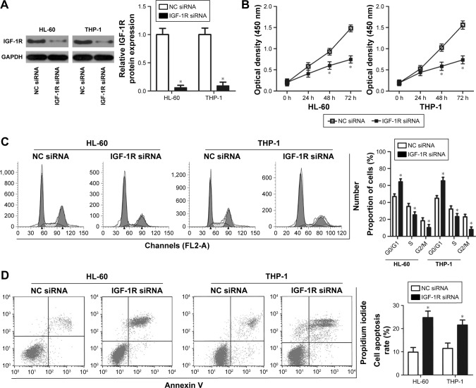
Background: A variety of microRNAs (miRNAs) are aberrantly expressed in acute myeloid leukemia (AML), and these dysregulated miRNAs perform crucial roles in tumorigenesis and progression of AML. miR-628-3p (miR-628), one of the miRNAs dysregulated in multiple types of human cancers, exerts antitumor roles in different cancer types. However, no specific study has explored the expression pattern and role of miR-628 in AML.
Materials and methods: In this study, RT-qPCR was performed to detect miR-628 expression in AML tissues and cell lines. CCK-8 assay, flow cytometry analysis and xenograft tumor experiment was carried out to determine the functions of miR-628 in AML cells. The possible mechanism underlying the activity of miR-628 in AML cells was also explored using a series of experiments.
Results: Our results revealed the downregulated expression of miR-628 in patients with AML and AML cell lines. Ectopic expression of miR-628 resulted in the inhibition of AML cell proliferation and induction of cell cycle arrest and apoptosis in vitro and attenuation of tumor growth in vivo. Insulin-like growth factor 1 receptor (IGF-1R) was identified as a direct target gene of miR-628 in AML cells. IGF-1R expression was upregulated in patients with AML and upregulation of IGF-1R expression inversely correlated with miR-628 level. Furthermore, IGF-1R knockdown imitated the tumor suppressive effect of miR-628 in AML cells. Restoration of IGF-1R expression abrogated the effects of miR-628 on the proliferation, cycle status, and apoptosis rate of AML cells. miR-628 inhibited the activation of phosphatidylinositol-4,5-bisphosphate 3-kinase (PI3K)/protein kinase B (Akt) pathway in AML cells both in vitro and in vivo through the inhibition of IGF-1R expression.
Conclusion: Our results demonstrate that miR-628 exhibits antitumor effects in AML through the direct targeting of IGF-1R and regulation of PI3K/Akt pathway, suggestive of its potential role as a therapeutic target in patients with this aggressive hematological malignant tumor.
Keywords: PI3K/Akt pathway; acute myeloid leukemia; apoptosis; cell cycle; insulin-like growth factor 1 receptor; microRNA-628; proliferation.
Conflict of interest statement
Disclosure The authors report no conflicts of interest in this work.
Figures


Similar articles
-
miR-338-3p Plays a Significant Role in Casticin-Induced Suppression of Acute Myeloid Leukemia via Targeting PI3K/Akt Pathway.Biomed Res Int. 2022 Jun 18;2022:9214130. doi: 10.1155/2022/9214130. eCollection 2022. Biomed Res Int. 2022. PMID: 35765408 Free PMC article.
-
microRNA-944 inhibits the malignancy of hepatocellular carcinoma by directly targeting IGF-1R and deactivating the PI3K/Akt signaling pathway.Cancer Manag Res. 2019 Mar 29;11:2531-2543. doi: 10.2147/CMAR.S199818. eCollection 2019. Cancer Manag Res. 2019. Retraction in: Cancer Manag Res. 2021 Jun 16;13:4765. doi: 10.2147/CMAR.S324204. PMID: 31114322 Free PMC article. Retracted.
-
microRNA-532 suppresses the PI3K/Akt signaling pathway to inhibit colorectal cancer progression by directly targeting IGF-1R.Am J Cancer Res. 2018 Mar 1;8(3):435-449. eCollection 2018. Am J Cancer Res. 2018. Retraction in: Am J Cancer Res. 2021 Sep 15;11(9):4641. PMID: 29636999 Free PMC article. Retracted.
-
Clinical Applications of MicroRNAs in Acute Myeloid Leukemia: A Mini-Review.Front Oncol. 2021 Aug 11;11:679022. doi: 10.3389/fonc.2021.679022. eCollection 2021. Front Oncol. 2021. PMID: 34458136 Free PMC article. Review.
-
IGF/IGF-1R signal pathway in pain: a promising therapeutic target.Int J Biol Sci. 2023 Jul 3;19(11):3472-3482. doi: 10.7150/ijbs.84353. eCollection 2023. Int J Biol Sci. 2023. PMID: 37497005 Free PMC article. Review.
Cited by
-
Homo sapiens circular RNA 0003602 (Hsa_circ_0003602) accelerates the tumorigenicity of acute myeloid leukemia by modulating miR-502-5p/IGF1R axis.Mol Cell Biochem. 2022 Feb;477(2):635-644. doi: 10.1007/s11010-021-04277-0. Epub 2022 Jan 6. Mol Cell Biochem. 2022. PMID: 34988853
-
miR-4792 Inhibits Acute Myeloid Leukemia Cell Proliferation and Invasion and Promotes Cell Apoptosis by Targeting Kindlin-3.Oncol Res. 2020 Sep 1;28(4):357-369. doi: 10.3727/096504020X15844389264424. Epub 2020 Mar 17. Oncol Res. 2020. PMID: 32183929 Free PMC article.
-
Comparison of microRNA Expression Profile in Chronic Myeloid Leukemia Patients Newly Diagnosed and Treated by Allogeneic Hematopoietic Stem Cell Transplantation.Front Oncol. 2020 Sep 4;10:1544. doi: 10.3389/fonc.2020.01544. eCollection 2020. Front Oncol. 2020. PMID: 33014798 Free PMC article.
-
MicroRNA‑628‑5p inhibits cell proliferation and induces apoptosis in colorectal cancer through downregulating CCND1 expression levels.Mol Med Rep. 2020 Mar;21(3):1481-1490. doi: 10.3892/mmr.2020.10945. Epub 2020 Jan 16. Mol Med Rep. 2020. Retraction in: Mol Med Rep. 2023 Sep;28(3):158. doi: 10.3892/mmr.2023.13045. PMID: 32016467 Free PMC article. Retracted.
-
MicroRNA-939-5p directly targets IGF-1R to inhibit the aggressive phenotypes of osteosarcoma through deactivating the PI3K/Akt pathway.Int J Mol Med. 2019 Nov;44(5):1833-1843. doi: 10.3892/ijmm.2019.4333. Epub 2019 Sep 5. Int J Mol Med. 2019. Retraction in: Int J Mol Med. 2022 Oct;50(4):125. doi: 10.3892/ijmm.2022.5181. PMID: 31545400 Free PMC article. Retracted.
References
-
- Short NJ, Ravandi F. Acute myeloid leukemia: past, present, and prospects for the future. Clin Lymphoma Myeloma Leuk. 2016;16(Suppl):S25–S29. - PubMed
-
- Schlenk RF, Dohner K, Krauter J. German-Austrian acute myeloid Leukemia Study. Mutations and treatment outcome in cytogenetically normal acute myeloid leukemia. N Engl J Med. 2008;358:1909–1918. - PubMed
-
- Woods BA, Levine RL. The role of mutations in epigenetic regulators in myeloid malignancies. Immunol Rev. 2015;263(1):22–35. - PubMed
Publication types
LinkOut - more resources
Full Text Sources
