

Transcatheter arterial embolization combined with hypoxia-replicative oncolytic adenovirus perfusion enhances the therapeutic effect of hepatic carcinoma
- PMID: 30774426
- PMCID: PMC6350642
- DOI: 10.2147/CMAR.S189208
Transcatheter arterial embolization combined with hypoxia-replicative oncolytic adenovirus perfusion enhances the therapeutic effect of hepatic carcinoma
Abstract
Purpose: Transcatheter arterial embolization or transcatheter arterial chemoembolization has become a critical therapy for unresectable hepatocarcinoma. Although hypoxia caused by embolization can induce apoptosis and necrosis of the majority of tumor cells, a small proportion of cells can survive with hypoxia and chemotherapy resistance. HIF-1α induced by hypoxia is the key factor rendering surviving tumor cells invasive and metastatic properties. Thus, we generated a synthetic hypoxia-replicative oncolytic adenovirus (HYAD) expecting to further eliminate the surviving tumor cells, which expressed HIF-1α.
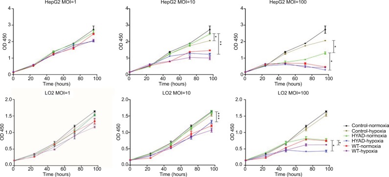
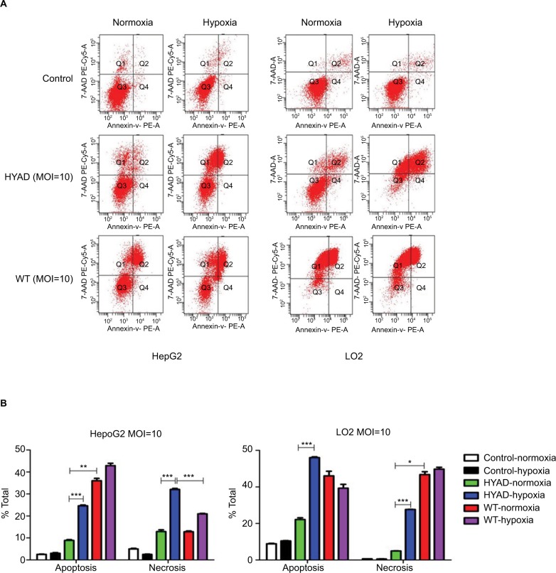
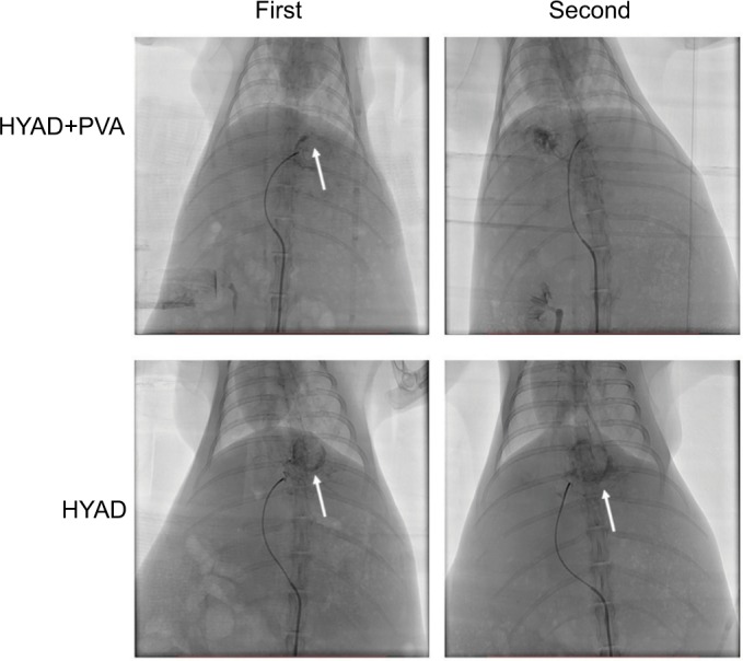
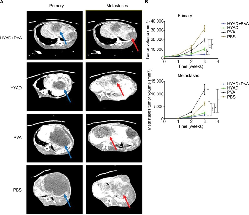
Materials and methods: In our study, we detected protein expression, proliferation, apoptosis, and necrosis of hepatic tumor cell line when infected with HYAD under hypoxia and normoxia. And we constructed VX2 hepatic cancer rabbit models to explore the therapeutic effect of transcatheter arterial embolization combined with HYAD perfusion under digital subtraction angiography. Inhibition of tumor growth and invasion was detected by histopathological examination and contrast-enhanced CT scan.
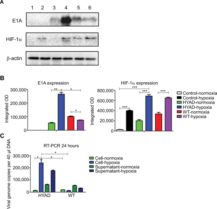
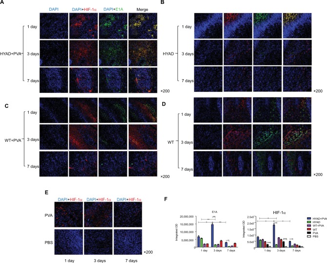
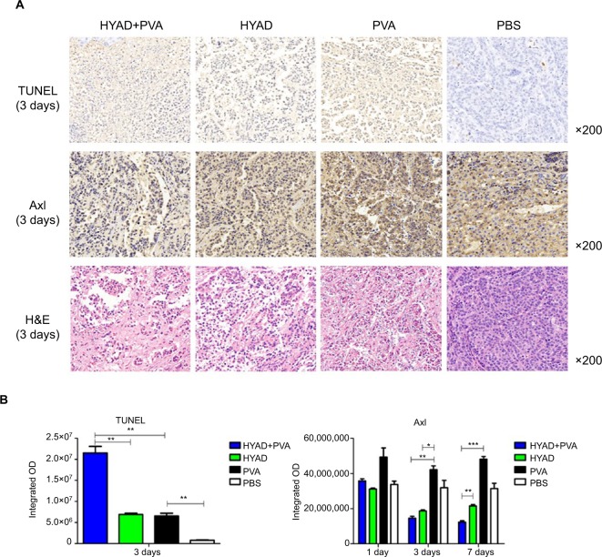
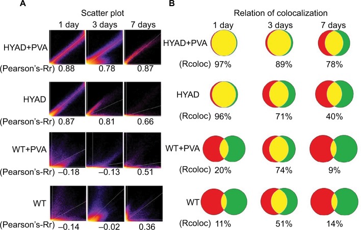
Results: Experiments in vitro verified that HYAD expressed and replicated along with HIF-1α expression or hypoxia. Compared with wild adenovirus type 5 (WT), HYAD expressed much more under hypoxia, which was the main principle of HYAD killing surviving tumor cells posttransarterial embolization. In vivo experiment of VX2 models, HYAD perfusion combined with polyvinyl alcohol (PVA) embolization achieved the highest expression quantity and the longest expression duration compared with simple HYAD perfusion, WT perfusion combined with PVA embolization, and simple WT perfusion. Because adenovirus expression protein E1A had the properties of promoting apoptosis, inhibiting invasion, and inhibiting metastasis, HYAD perfusion combined with PVA embolization group efficiently repressed tumor growth and intrahepatic metastases compared to other processing groups.
Conclusion: HYAD can overcome the hypoxic tumor microenvironment postembolization and target the surviving tumor cells with specificity. In turn, HYAD perfusion combined with PVA embolization can bring out the best effect in each other.
Keywords: hepatocarcinoma; hypoxia; oncolytic adenovirus; transcatheter arterial embolization.
Conflict of interest statement
Disclosure The authors report no conflicts of interest in this work.
Figures
Similar articles
-
Hepatocellular necrosis, apoptosis, and proliferation after transcatheter arterial embolization or chemoembolization in a standardized rabbit model.J Vasc Interv Radiol. 2011 Nov;22(11):1606-12. doi: 10.1016/j.jvir.2011.08.005. Epub 2011 Sep 29. J Vasc Interv Radiol. 2011. PMID: 21959058
-
Antitumor effect of curcumin liposome after transcatheter arterial embolization in VX2 rabbits.Cancer Biol Ther. 2019;20(5):642-652. doi: 10.1080/15384047.2018.1550567. Epub 2019 Jan 8. Cancer Biol Ther. 2019. PMID: 30621501 Free PMC article.
-
Liposomal curcumin inhibits hypoxia-induced angiogenesis after transcatheter arterial embolization in VX2 rabbit liver tumors.Onco Targets Ther. 2015 Sep 15;8:2601-11. doi: 10.2147/OTT.S87931. eCollection 2015. Onco Targets Ther. 2015. PMID: 26451117 Free PMC article.
-
Effect of transcatheter arterial embolization on levels of hypoxia-inducible factor-1alpha in rabbit VX2 liver tumors.J Vasc Interv Radiol. 2007 May;18(5):639-45. doi: 10.1016/j.jvir.2007.02.031. J Vasc Interv Radiol. 2007. PMID: 17494846
-
Experimental evaluation of inhibitory effect of 10-hydroxycamptothecin on hypoxia-inducible factor-1α expression and angiogenesis in liver tumors after transcatheter arterial embolization.J Vasc Interv Radiol. 2010 Oct;21(10):1565-72. doi: 10.1016/j.jvir.2010.05.028. Epub 2010 Sep 1. J Vasc Interv Radiol. 2010. PMID: 20810290
Cited by
-
Postoperative Survival and Clinical Outcomes for Uterine Leiomyosarcoma Spinal Bone Metastasis: A Case Series and Systematic Literature Review.Diagnostics (Basel). 2022 Dec 21;13(1):15. doi: 10.3390/diagnostics13010015. Diagnostics (Basel). 2022. PMID: 36611309 Free PMC article.
-
Targeting Hypoxia-Inducible Factor-1α for the Management of Hepatocellular Carcinoma.Cancers (Basel). 2023 May 12;15(10):2738. doi: 10.3390/cancers15102738. Cancers (Basel). 2023. PMID: 37345074 Free PMC article. Review.
-
Hypoxia as a Target for Combination with Transarterial Chemoembolization in Hepatocellular Carcinoma.Pharmaceuticals (Basel). 2024 Aug 11;17(8):1057. doi: 10.3390/ph17081057. Pharmaceuticals (Basel). 2024. PMID: 39204162 Free PMC article. Review.
-
Oncolytic virus therapy in hepatocellular carcinoma.Oncol Res. 2025 Jun 26;33(7):1593-1610. doi: 10.32604/or.2025.061857. eCollection 2025. Oncol Res. 2025. PMID: 40612869 Free PMC article. Review.
References
-
- Heimbach JK, Kulik LM, Finn RS, et al. AASLD guidelines for the treatment of hepatocellular carcinoma. Hepatology. 2018;67(1):358–380. - PubMed
-
- Song P, Cai Y, Tang H, Li C, Huang J. The clinical management of hepatocellular carcinoma worldwide: a concise review and comparison of current guidelines from 2001 to 2017. Biosci Trends. 2017;11(4):389–398. - PubMed
-
- Erhardt A, Kolligs F, Dollinger M, et al. TACE plus sorafenib for the treatment of hepatocellular carcinoma: results of the multi-center, phase II SOCRATES trial. Cancer Chemother Pharmacol. 2014;74(5):947–954. - PubMed
LinkOut - more resources
Full Text Sources
Miscellaneous
