Carbon-ion radiotherapy in accelerated hypofractionated active raster-scanning technique for malignant lacrimal gland tumors: feasibility and safety
- PMID: 30774443
- PMCID: PMC6362930
- DOI: 10.2147/CMAR.S190051
Carbon-ion radiotherapy in accelerated hypofractionated active raster-scanning technique for malignant lacrimal gland tumors: feasibility and safety
Abstract
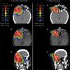
Introduction: We evaluated treatment outcomes of CIRT in an active raster-scanning technique alone or in combination with IMRT for lacrimal gland tumors.
Methods: A total of 24 patients who received CIRT for a malignant lacrimal gland tumor at the HIT between 2009 and 2018 were analyzed retrospectively for LC, OS, and distant progression-free survival (DPFS) using Kaplan-Meier estimates. Toxicity was assessed according to the CTCAE version 5.
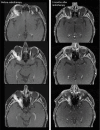
Results: Median follow-up was 30 months and overall median LC, OS, and DPFS 24 months, 36 months, and 31 months, respectively. Two-year LC, OS, and DPFS of 93%, 96%, and 87% with CIRT was achieved for all patients. Local failure occurred only in patients with ACC and after a median follow-up of 30 months after the completion of RT (n=5, 21%; P=0.09). We identified a significant negative impact of a macroscopic tumor disease, which was diagnosed on planning CT or MRI before RT, on LC (P=0.026). In contrast, perineural spread (P=0.661), T stage (P=0.552), and resection margins in operated patients (P=0.069) had no significant impact on LC. No grade ≥3 acute or grade >3 chronic toxicity occurred. Late grade 3 side effects were identified in form of a wound-healing disorder 3 months after RT in one patient and temporal lobe necrosis 6 months after RT in another (n=2, 8%).
Conclusion: Accelerated hypofractionated active raster-scanning CIRT for relative radio-resistant malignant lacrimal gland tumors results in adequate LC rates and moderate acute and late toxicity. Nevertheless, LC for ACC histology remains challenging and risk factors for local recurrence are still unclear. Further follow-up is necessary to evaluate long-term clinical outcome.
Keywords: adenoid cystic carcinoma; bimodal RT; carbon-ion radiotherapy; local control; malignant lacrimal gland tumor.
Conflict of interest statement
Disclosure The authors report no conflicts of interest in this work.
Figures


References
-
- Shields JA, Shields CL, Epstein JA, Scartozzi R, Eagle RC. Review: primary epithelial malignancies of the lacrimal gland: the 2003 Ramon L. Font lecture. Ophthalmic Plast Reconstr Surg. 2004;20(1):10–21. - PubMed
-
- von Holstein SL, Therkildsen MH, Prause JU, Stenman G, Siersma VD, Heegaard S. Lacrimal gland lesions in Denmark between 1974 and 2007. Acta Ophthalmol. 2013;91(4):349–354. - PubMed
-
- Cl S, Shields JA. Review of lacrimal gland lesions. Trans Pa Acad Ophthalmol Otolaryngol. 1990;42:925–930. - PubMed
-
- Gao Y, Moonis G, Cunnane ME, Eisenberg RL. Lacrimal gland masses. AJR Am J Roentgenol. 2013;201(3):W371–W381. - PubMed
-
- Ni C, Kuo PK, Dryja TP. Histopathological classification of 272 primary epithelial tumors of the lacrimal gland. Chin Med J (Engl) 1992;105(6):481–485. - PubMed
LinkOut - more resources
Full Text Sources

