

Management of foot and ankle injuries in pediatric and adolescent athletes: a narrative review
- PMID: 30774457
- PMCID: PMC6209353
- DOI: 10.2147/ORR.S129990
Management of foot and ankle injuries in pediatric and adolescent athletes: a narrative review
Abstract
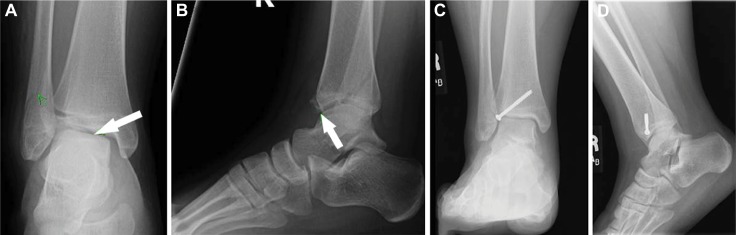
In this review, we focus on the treatment of injuries to the foot and ankle in the adolescent athlete. While many injuries in the adolescent foot and ankle are similar to or overlap with their counterparts in the adult population, the anatomy of the adolescent ankle, especially the presence of growth plates, results in different injury patterns in many cases and calls for specific management approaches. We discuss the unique anatomy of the pediatric patient as well as the diagnostic evaluation and treatment of common injuries in the young athlete.
Keywords: Lisfranc injury; ankle; foot; growth plate; lower extremity; pediatric athlete.
Conflict of interest statement
Disclosure The authors received no funding for this study, and report no conflicts of interest.
Figures
Similar articles
-
Common injuries of the foot and ankle in the child and adolescent athlete.Phys Med Rehabil Clin N Am. 2008 May;19(2):347-71, ix. doi: 10.1016/j.pmr.2007.11.003. Phys Med Rehabil Clin N Am. 2008. PMID: 18395652 Review.
-
Ankle Injuries in the Pediatric Athlete.Sports Med Arthrosc Rev. 2016 Dec;24(4):170-177. doi: 10.1097/JSA.0000000000000125. Sports Med Arthrosc Rev. 2016. PMID: 27811516 Review.
-
Ankle and Foot Injuries in the Young Athlete.Semin Musculoskelet Radiol. 2018 Feb;22(1):104-117. doi: 10.1055/s-0037-1609012. Epub 2018 Feb 6. Semin Musculoskelet Radiol. 2018. PMID: 29409077 Review.
-
Physical therapy and rehabilitation of the foot and ankle in the athlete.Clin Podiatr Med Surg. 2011 Jan;28(1):189-201. doi: 10.1016/j.cpm.2010.09.005. Clin Podiatr Med Surg. 2011. PMID: 21276526 Review.
-
Ankle and foot injuries in pediatric and adult athletes.Prim Care. 2005 Mar;32(1):133-61. doi: 10.1016/j.pop.2004.11.003. Prim Care. 2005. PMID: 15831316 Review.
Cited by
-
Age- and gender-specific magnetic resonance imaging findings in paediatric ankle trauma with negative radiographs.BMC Musculoskelet Disord. 2025 Sep 15;26(1):851. doi: 10.1186/s12891-025-09155-0. BMC Musculoskelet Disord. 2025. PMID: 40954486 No abstract available.
-
Addressing the Need for a Better Understanding of Foot and Ankle Pathology Relevant to the Adolescent Athlete.Children (Basel). 2023 Aug 3;10(8):1342. doi: 10.3390/children10081342. Children (Basel). 2023. PMID: 37628340 Free PMC article.
-
Treatment Variability and Complications Associated With Pediatric Lateral Ankle Injuries: A POSNA Quality, Safety, and Value Initiative Survey.Orthop J Sports Med. 2022 Jun 1;10(6):23259671221100223. doi: 10.1177/23259671221100223. eCollection 2022 Jun. Orthop J Sports Med. 2022. PMID: 35668871 Free PMC article.
-
Progress in diagnosis and treatment of acute injury to the anterior talofibular ligament.World J Clin Cases. 2023 May 26;11(15):3395-3407. doi: 10.12998/wjcc.v11.i15.3395. World J Clin Cases. 2023. PMID: 37383912 Free PMC article. Review.
-
Magnetic Resonance Imaging Patterns of Common Injuries in Pediatric and Adolescent Athletes.HSS J. 2024 Aug;20(3):390-401. doi: 10.1177/15563316241233578. Epub 2024 Mar 28. HSS J. 2024. PMID: 39108444 Free PMC article.
References
-
- Kay RM, Matthys GA. Pediatric ankle fractures: evaluation and treatment. J Am Acad Orthop Surg. 2001;9(4):268–278. - PubMed
-
- Herring JA. Tachdjian’s Pediatric Orthopaedics: From the Texas Scottish Rite Hospital for Children. 5th ed. New York: Elsevier Saunders; 2013.
-
- Ogden JA, Lee J. Accessory ossification patterns and injuries of the malleoli. J Pediatr Orthop. 1990;10(3):306–316. - PubMed
-
- Canale ST, Williams KD. Iselin’s disease. J Pediatr Orthop. 1992;12(1):90–93. - PubMed
-
- Lawson JP. Symptomatic radiographic variants in extremities. Radiology. 1985;157(3):625–631. - PubMed
Publication types
LinkOut - more resources
Full Text Sources
