

The etiology, evaluation, and management of plantar fibromatosis
- PMID: 30774465
- PMCID: PMC6367723
- DOI: 10.2147/ORR.S154289
The etiology, evaluation, and management of plantar fibromatosis
Abstract
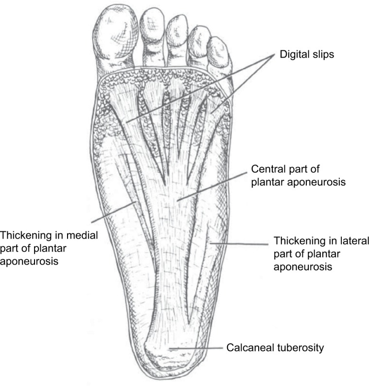
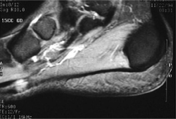
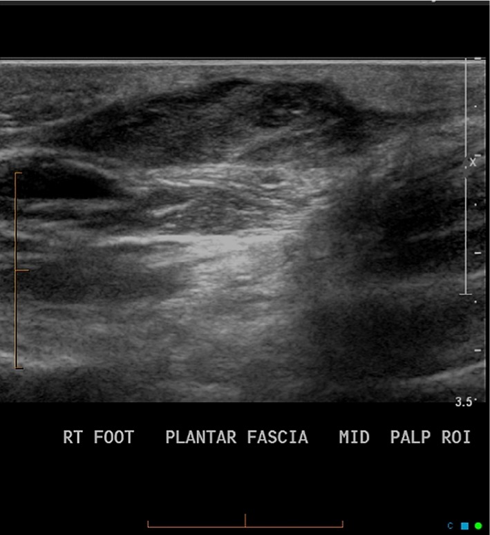
Plantar fibromatosis (Ledderhose disease) is a rare, benign, hyperproliferative fibrous tissue disorder resulting in the formation of nodules along the plantar fascia. This condition can be locally aggressive, and often results in pain, functional disability, and decreased quality of life. Diagnosis is primarily clinical, but MRI and ultrasound are useful confirmatory adjuncts. Given the benign nature of this condition, treatment has historically involved symptomatic management. A multitude of conservative treatment strategies supported by varying levels of evidence have been described mostly in small-scale trials. These therapies include steroid injections, verapamil, radiation therapy, extracorporeal shock wave therapy, tamoxifen, and collagenase. When conservative measures fail, surgical removal of fibromas and adjacent plantar fascia is often done, although recurrence is common. This review aims to provide a broad overview of the clinical features of this disease as well as the current treatment strategies being employed in the management of this condition.
Keywords: Ledderhose disease; plantar fascia; plantar fibromatosis.
Conflict of interest statement
Disclosure All authors report no conflicts of interest in this work.
Figures
References
-
- Lee TH, Wapner KL, Hecht PJ. Plantar fibromatosis. J Bone Joint Surg Am. 1993;75(7):1080–1084. - PubMed
-
- Dürr HR, Krödel A, Trouillier H, Lienemann A, Refior HJ. Fibromatosis of the plantar fascia: diagnosis and indications for surgical treatment. Foot Ankle Int. 1999;20(1):13–17. - PubMed
-
- Rosenbaum AJ, Dipreta JA, Misener D. Plantar heel pain. Med Clin North Am. 2014;98(2):339–352. - PubMed
-
- Lareau CR, Sawyer GA, Wang JH, Digiovanni CW. Plantar and medial heel pain: diagnosis and management. J Am Acad Orthop Surg. 2014;22(6):372–380. - PubMed
-
- Neufeld SK, Cerrato R. Plantar fasciitis: evaluation and treatment. J Am Acad Orthop Surg. 2008;16(6):338–346. - PubMed
Publication types
LinkOut - more resources
Full Text Sources
