

3D-printed patient-specific applications in orthopedics
- PMID: 30774470
- PMCID: PMC6209352
- DOI: 10.2147/ORR.S99614
3D-printed patient-specific applications in orthopedics
Abstract
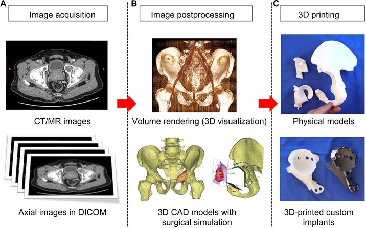
With advances in both medical imaging and computer programming, two-dimensional axial images can be processed into other reformatted views (sagittal and coronal) and three-dimensional (3D) virtual models that represent a patients' own anatomy. This processed digital information can be analyzed in detail by orthopedic surgeons to perform patient-specific orthopedic procedures. The use of 3D printing is rising and has become more prevalent in medical applications over the last decade as surgeons and researchers are increasingly utilizing the technology's flexibility in manufacturing objects. 3D printing is a type of manufacturing process in which materials such as plastic or metal are deposited in layers to create a 3D object from a digital model. This additive manufacturing method has the advantage of fabricating objects with complex freeform geometry, which is impossible using traditional subtractive manufacturing methods. Specifically in surgical applications, the 3D printing techniques can not only generate models that give a better understanding of the complex anatomy and pathology of the patients and aid in education and surgical training, but can also produce patient-specific surgical guides or even custom implants that are tailor-made to the surgical requirements. As the clinical workflow of the 3D printing technology continues to evolve, orthopedic surgeons should embrace the latest knowledge of the technology and incorporate it into their clinical practice for patient-specific orthopedic applications. This paper is written to help orthopedic surgeons stay up-to-date on the emerging 3D technology, starting from the acquisition of clinical imaging to 3D printing for patient-specific applications in orthopedics. It 1) presents the necessary steps to prepare the medical images that are required for 3D printing, 2) reviews the current applications of 3D printing in patient-specific orthopedic procedures, 3) discusses the potential advantages and limitations of 3D-printed custom orthopedic implants, and 4) suggests the directions for future development. The 3D printing technology has been reported to be beneficial in patient-specific orthopedics, such as in the creation of anatomic models for surgical planning, education and surgical training, patient-specific instruments, and 3D-printed custom implants. Besides being anatomically conformed to a patient's surgical requirement, 3D-printed implants can be fabricated with scaffold lattices that may facilitate osteointegration and reduce implant stiffness. However, limitations including high cost of the implants, the lead time in manufacturing, and lack of intraoperative flexibility need to be addressed. New biomimetic materials have been investigated for use in 3D printing. To increase utilization of 3D printing technology in orthopedics, an all-in-one computer platform should be developed for easy planning and seamless communications among different care providers. Further studies are needed to investigate the real clinical efficacy of 3D printings in orthopedic applications.
Keywords: 3D printing; custom implants; image processing; patient-specific instrument; patient-specific orthopedics.
Conflict of interest statement
Disclosure The Materialise, Mobelife company did not fund or sponsor this research in any way. The author reports no conflicts of interest in this work.
Figures


References
-
- Schubert C, Van Langeveld MC, Donoso LA. Innovations in 3D printing: a 3D overview from optics to organs. Br J Ophthalmol. 2014;98:159–161. - PubMed
-
- Martelli N, Serrano C, van den Brink H, et al. Advantages and disadvantages of 3-dimensional printing in surgery: a systematic review. Surgery. 2016;159(6):1485–1500. - PubMed
-
- Lu S, Xu YQ, Lu WW, et al. A novel patient-specific navigational template for cervical pedicle screw placement. Spine. 2009;34(26):E959–E966. - PubMed
Publication types
LinkOut - more resources
Full Text Sources
Other Literature Sources
Research Materials
